Pityriasis Rosea: An Overview
Introduction
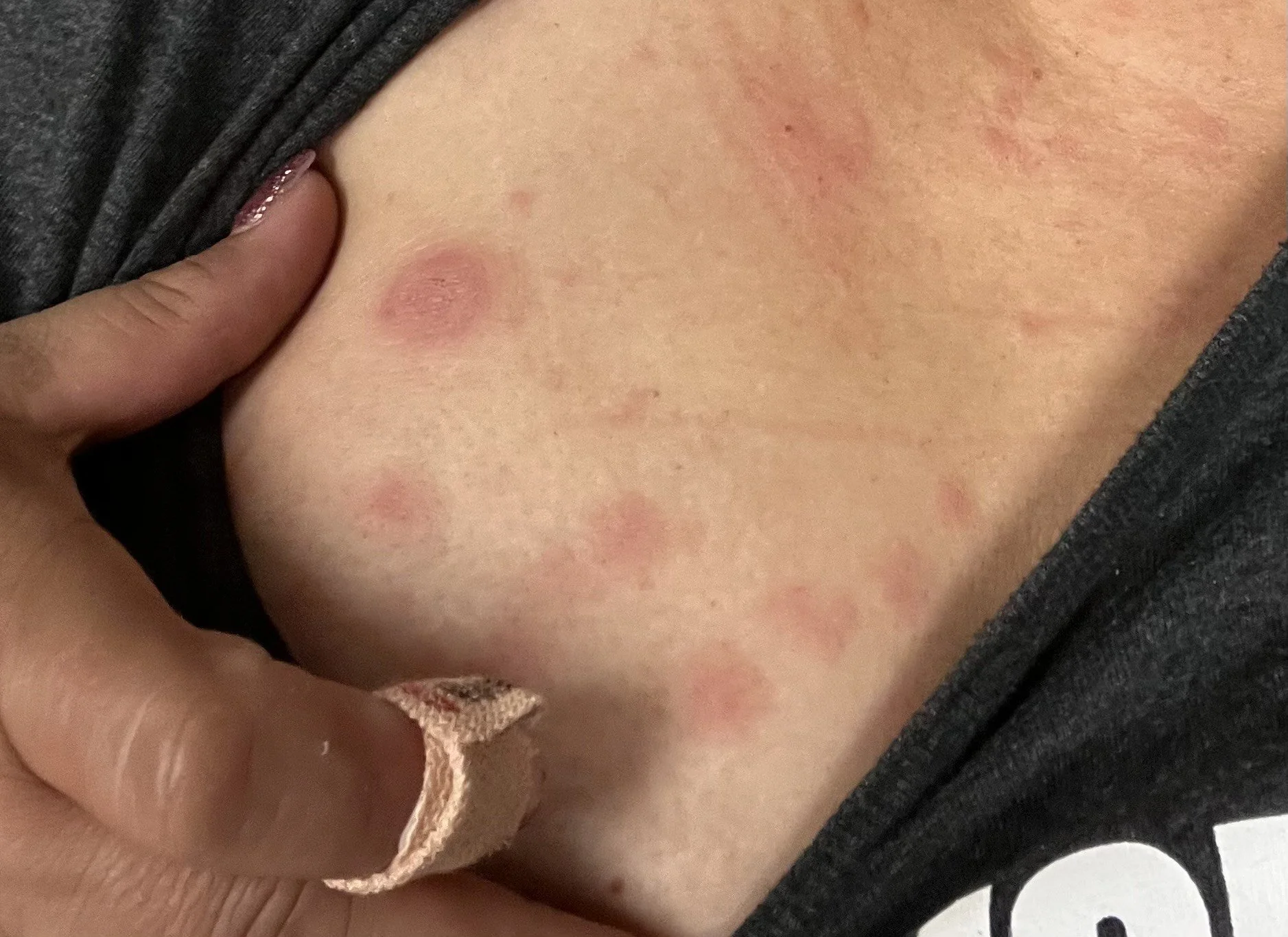
Pityriasis rosea (PR) is an acute, self-limiting skin disorder primarily affecting older children and young adults, with a slightly higher prevalence in females. This condition is marked by distinctive, oval-shaped, scaly lesions typically on the torso and upper limbs. PR is characterized by an initial "herald patch"—a solitary, round or oval lesion that appears on the chest, neck, or back. This initial lesion is often followed by multiple, smaller lesions that emerge in a characteristic "Christmas tree" pattern on the back, aligned with the skin’s cleavage lines. PR tends to resolve on its own, generally within two to three months, although it can persist longer in some cases.
Etiology and Pathogenesis
The exact cause of PR remains uncertain, though evidence suggests a possible viral origin. Several studies indicate an association between PR and human herpesviruses, particularly human herpesvirus 7 (HHV-7) and, to a lesser extent, HHV-6. The reactivation of these latent viruses is thought to trigger the skin reaction seen in PR. Further supporting the viral hypothesis, some PR cases are preceded by mild prodromal symptoms, such as headache, malaise, and sore throat, mimicking a viral infection. Additionally, PR is known to sometimes occur in clusters, suggesting potential environmental or viral spread, although it is not considered highly contagious.
The hypothesis of a viral etiology remains inconclusive due to conflicting research findings. Viral particles resembling herpesvirus have been detected in PR lesions in some studies, while others have failed to confirm the presence of HHV-7 or HHV-6 DNA in PR biopsy samples. Other viral agents, including HHV-8, influenza A (H1N1), and SARS-CoV-2, have also been suggested as possible contributors or triggers, especially following observations of PR-like eruptions in patients during the COVID-19 pandemic. However, these associations require further investigation.
Clinical Presentation
PR typically begins with the development of a herald patch, a single, round or oval lesion ranging from 2 to 5 cm in diameter. This lesion is well-defined, with a slightly scaly center that often clears, leaving a distinct border or "collarette" of scale around its edges. After the herald patch appears, smaller, similar lesions usually follow within one to two weeks, spreading over the trunk and proximal extremities in a pattern that resembles a fir or Christmas tree on the back.
Most PR cases are asymptomatic aside from mild itching, but some patients report prodromal symptoms like sore throat, headache, and malaise. The lesions generally fade within four to six weeks, though some individuals, especially those with darker skin tones, may experience post-inflammatory hyperpigmentation lasting several months. PR has a mild impact on quality of life, but extensive or prolonged cases may be more distressing, particularly in those with atypical presentations, such as facial or extremity involvement.
Differential Diagnosis
PR's presentation can mimic several dermatologic conditions, making an accurate differential diagnosis important. Skin biopsy and laboratory testing, though rarely required, may be performed in cases of diagnostic uncertainty. Key conditions to consider in the differential diagnosis include:
Secondary Syphilis: Like PR, secondary syphilis presents as a papulosquamous rash, often involving the trunk. However, secondary syphilis lesions frequently appear on the palms and soles, a feature uncommon in PR. A history of primary syphilis (chancre) or serologic testing can help confirm this diagnosis.
Guttate Psoriasis: This psoriasis subtype, often linked to a streptococcal infection, produces small, erythematous, scaly plaques on the trunk and limbs. Unlike PR, guttate psoriasis lacks a herald patch and typically presents with a more persistent scale.
Tinea Corporis: The herald patch of PR may resemble the annular plaques seen in tinea corporis. Potassium hydroxide (KOH) testing can help identify fungal elements in tinea corporis, confirming a dermatophyte infection if present.
Tinea Versicolor: This condition causes hypopigmented or hyperpigmented macules, particularly on the trunk and neck. Unlike PR, lesions in tinea versicolor generally lack erythema and have a fine scale without the characteristic collarette seen in PR.
Nummular Eczema and Pityriasis Lichenoides Chronica: Nummular eczema presents with coin-shaped plaques that are intensely pruritic, unlike the generally mild itching seen in PR. Pityriasis lichenoides chronica causes recurrent crops of red to brown scaly papules, with lesions persisting for extended periods.
Diagnosis
Diagnosing PR is largely clinical, relying on the presence of a herald patch, characteristic lesion morphology, and pattern distribution. The herald patch can closely resemble tinea corporis, so a KOH preparation test may be performed to rule out a fungal infection. In sexually active patients, syphilis should also be excluded, especially in cases with widespread or atypical lesions. Biopsy is typically unnecessary but, when performed, may reveal non-specific findings, such as parakeratosis, lymphocytic infiltration, and red cell extravasation.
Treatment and Management
PR is self-limiting and typically resolves without intervention, so patient education and reassurance are key components of management. Most cases require no medical treatment aside from pruritus relief, which can be achieved with medium-potency topical corticosteroids or antipruritic lotions containing pramoxine or menthol. Oral antihistamines may also be helpful for patients experiencing bothersome itching.
In severe cases, additional interventions may be considered. Oral acyclovir has been studied due to PR’s possible link to herpesviruses, with mixed results. Acyclovir appears to accelerate symptom resolution in some cases but is not routinely recommended due to limited data on efficacy. Phototherapy, particularly broadband or narrowband UVB, has shown potential benefits in reducing lesion severity and pruritus, although studies are limited, and exposure to sunlight can worsen PR in some individuals.
Macrolide antibiotics, such as erythromycin, have been investigated but are not widely recommended due to inconclusive evidence regarding their effectiveness. Systemic corticosteroids, though occasionally helpful for symptom control, are generally avoided due to concerns of potential relapse following discontinuation.
Prognosis and Long-Term Outlook
The prognosis for PR is favorable, with most cases resolving spontaneously within six to twelve weeks. While recurrences are rare, post-inflammatory hyperpigmentation may persist for several months in individuals with darker skin tones. Patient reassurance is essential, as PR is neither highly contagious nor indicative of a severe underlying condition.
PR and Pregnancy
Data on PR in pregnancy are limited, though some studies suggest an association with adverse outcomes, including an elevated risk of spontaneous abortion if PR occurs early in pregnancy. Further research is needed to determine the extent of this risk. Pregnant patients should consult their healthcare provider for guidance on managing PR symptoms.
Conclusion
Pityriasis rosea is an acute, self-limiting skin condition with characteristic lesions and distribution patterns. Although its exact cause remains unclear, PR is likely viral in origin, with links to human herpesviruses HHV-6 and HHV-7. Management primarily involves symptom relief, and most cases resolve within two to three months without intervention. Awareness of PR’s distinctive presentation can aid in accurate diagnosis and avoid unnecessary treatments, ensuring patients receive appropriate reassurance and care.

