
I Finished Accutane—Why Am I Still Breaking Out? Understanding Hormonal Acne After Isotretinoin
A 20-year-old woman continued to experience jawline acne after completing a full course of Accutane. Learn why hormonal acne can persist after isotretinoin, how tretinoin and birth control help maintain clear skin, and what treatment options are available at Village Dermatology in Houston and Katy, Texas.
Completing a course of Accutane® (isotretinoin) is a major milestone for patients with severe acne. Many people expect perfectly clear skin immediately after finishing treatment, so it can be frustrating when new pimples begin to appear only weeks later.
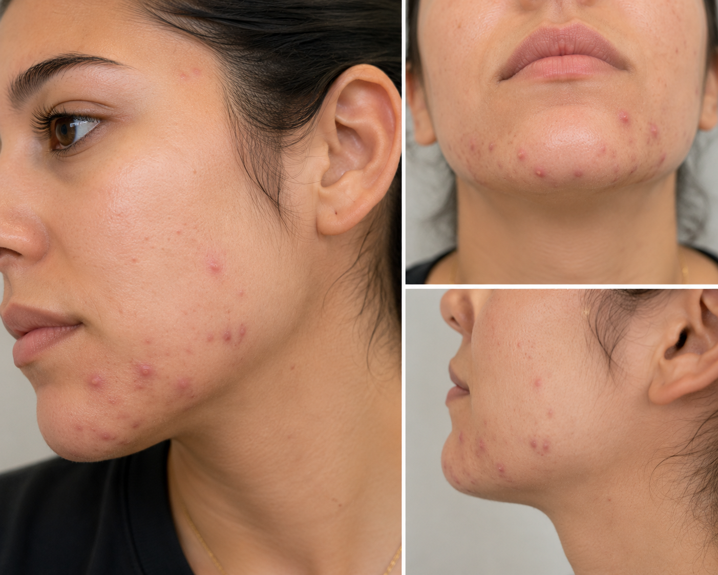
Recently, a 20-year-old woman visited our dermatology clinic after completing a full course of isotretinoin at another practice. Although her acne had improved dramatically, she continued to experience breakouts along her jawline. Rather than restarting Accutane, our board-certified dermatologist developed a personalized maintenance plan to help keep her skin clear while addressing the hormonal component of her acne.
If you've recently finished Accutane and are noticing new breakouts, here's what you should know.
The Patient's Story
This patient presented for evaluation approximately two weeks after completing a full course of isotretinoin.
She shared that:
She reached her target cumulative dose of Accutane.
Her acne improved significantly compared to where it started.
She was still developing occasional inflammatory breakouts.
The pimples were concentrated along her jawline, a common location for hormonal acne.
She had also started Lo Loestrin Fe, an oral contraceptive, during her isotretinoin treatment and had continued taking it after finishing therapy.
Additionally, she reported:
One month of irregular menstrual cycles after starting birth control.
Significant stress while beginning a new internship.
Concern that possible mold exposure in her home may have contributed to her previous severe acne flare, although the exact cause remained uncertain.
After examining her skin, the dermatologist determined that her remaining acne was most consistent with hormonal acne rather than treatment failure.
Can Acne Return After Accutane?
Yes.
Although isotretinoin is the most effective medication available for severe acne, some patients continue to develop mild breakouts after completing treatment, particularly if hormonal factors are involved.
This does not necessarily mean Accutane failed.
Instead, it often reflects ongoing hormonal stimulation of the oil glands.
Patients most likely to experience post-Accutane hormonal acne include those with:
Jawline acne
Menstrual-related flares
Adult female acne
Family history of persistent acne
Fortunately, maintenance therapy can often keep these breakouts under excellent control.
Why Is Jawline Acne Often Hormonal?
Acne that develops primarily along the:
Chin
Jawline
Lower cheeks
Neck
is commonly influenced by hormones called androgens.
These hormones stimulate the sebaceous (oil) glands, increasing oil production and creating an environment where acne develops more easily.
Stress, hormonal fluctuations, and genetics can all contribute to these breakouts.
Why Continue Birth Control After Accutane?
Combination oral contraceptives such as Lo Loestrin Fe can be an effective long-term treatment for hormonal acne.
They work by:
Lowering free testosterone levels
Reducing oil production
Helping prevent future hormonal breakouts
Patients should know that birth control pills often require two to three months before significant acne improvement becomes noticeable.
Irregular bleeding or menstrual changes may occur during the first few months as the body adjusts.
Why Was Tretinoin Started?
Rather than restarting isotretinoin, the patient transitioned to tretinoin 0.025% cream, a topical retinoid that serves as an excellent maintenance treatment.
Tretinoin helps by:
Preventing clogged pores
Increasing healthy skin cell turnover
Reducing new acne lesions
Improving skin texture over time
Gradually softening post-acne discoloration
To minimize irritation, she was instructed to:
Apply a pea-sized amount to the entire face.
Start 2–3 nights per week.
Increase to nightly use as tolerated.
Follow each application with a gentle moisturizer.
Gentle Skin Care Still Matters
Even after Accutane, maintaining a simple skincare routine is essential.
Patients are encouraged to use:
Gentle, non-comedogenic cleansers
Oil-free moisturizers
Broad-spectrum SPF 30+ sunscreen every day
Non-comedogenic makeup and skincare products
Avoiding harsh scrubs and over-cleansing can help preserve the skin barrier and reduce irritation from tretinoin.
What About Acne Scarring?
In addition to active acne, this patient also had acne scarring, which can persist long after breakouts improve.
Acne scars develop when deep inflammation damages the skin's collagen during healing.
While scars are permanent, their appearance can often be significantly improved with treatment.
Depending on the type of scarring, options may include:
Fractional laser resurfacing
Microneedling
Chemical peels
Dermabrasion
Combination laser therapies for redness and texture
Many scars also continue to soften naturally during the first one to two years after acne becomes controlled.
When Should You Return to Your Dermatologist?
Follow-up is recommended if:
Breakouts become more frequent.
Painful cysts or nodules return.
New acne scars develop.
Medications cause irritation or side effects.
Acne fails to improve after several months of maintenance therapy.
Early adjustments can often prevent future scarring and reduce the need for another course of isotretinoin.
Hormonal Acne Treatment in Houston and Katy, Texas
Finishing Accutane is an important achievement, but maintaining clear skin often requires a long-term strategy. Hormonal acne is one of the most common reasons patients continue to experience occasional breakouts after isotretinoin, and maintenance treatments such as topical retinoids and hormonal therapy can help preserve your results.
At Village Dermatology, our board-certified dermatologists provide personalized treatment for hormonal acne, acne scarring, and post-isotretinoin maintenance. We proudly care for patients throughout Houston and Katy, Texas, offering comprehensive acne treatment plans tailored to each patient's skin and lifestyle.
If you're still breaking out after Accutane or are looking for expert acne care, schedule an appointment with Village Dermatology to discuss your options.
Persistent Groin Rash Despite Nystatin? It May Be Intertrigo, Not Just a Yeast Infection
A 75-year-old woman developed a chronic groin and buttock rash that persisted despite nystatin treatment. Learn how dermatologists diagnose intertrigo, why barrier creams are essential, and when a medication-related rash should be evaluated at Village Dermatology in Houston and Katy, Texas.
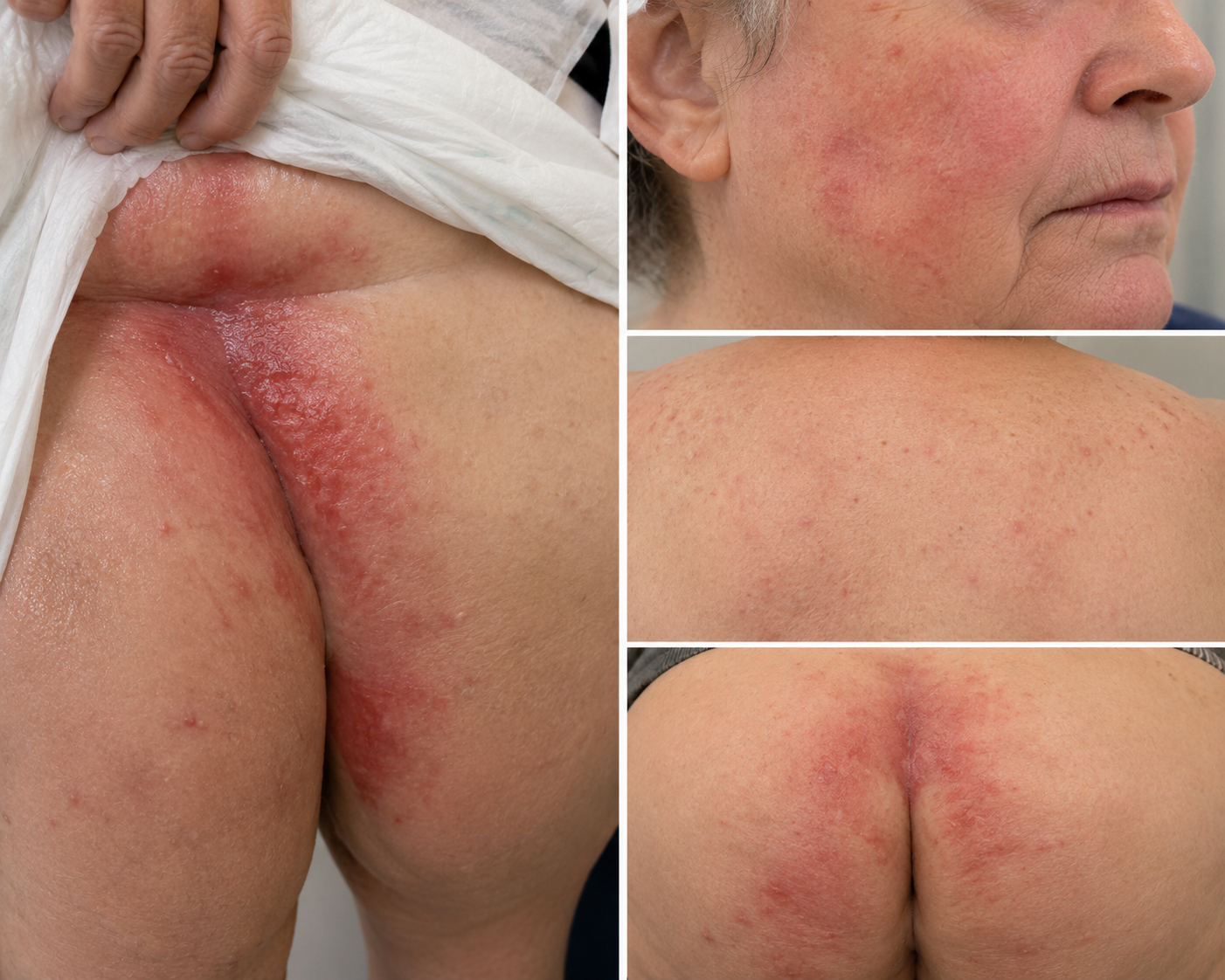
A persistent rash in the groin or buttocks can be uncomfortable, painful, and difficult to treat—especially in older adults who wear protective briefs or experience urinary or fecal incontinence. While many people assume these rashes are simply yeast infections, moisture, friction, and skin irritation often play equally important roles.
Recently, a 75-year-old woman visited our dermatology clinic with two separate skin concerns: a chronic rash affecting the groin and buttocks and a newer rash involving the face and back that developed after starting a new cholesterol medication.
After a thorough skin examination, our board-certified dermatologist diagnosed intertrigo with irritant contact dermatitis (ICD) in the skin folds and evaluated the second rash as a possible drug eruption versus dermatitis.
If you're searching for answers about persistent skin fold rashes or medication-related rashes in Houston or Katy, Texas, here's what you should know.
The Patient's Story
The patient was accompanied by her daughter, who explained that her mother had experienced recurring redness and irritation in the:
Groin
Buttocks
Gluteal cleft
Upper inner thigh
The rash had been present for several months and repeatedly flared despite treatment with nystatin ointment prescribed elsewhere.
She also developed a separate rash affecting the:
Face
Back
Shortly after starting atorvastatin, raising concern that the medication may have contributed.
Why Didn't the Nystatin Alone Work?
During the examination, the dermatologist observed:
Weepy skin
Maceration (softened skin caused by excess moisture)
Bright red inflamed patches
These findings suggested intertrigo, a condition caused by:
Moisture
Skin-on-skin friction
Heat
Irritation
Rather than a simple yeast infection alone.
Because the patient wore adult diapers and frequently had prolonged exposure to urine and stool, irritant contact dermatitis was also contributing to the skin breakdown.
In these situations, treating only yeast often fails because the underlying moisture and irritation remain.
What Is Intertrigo?
Intertrigo is an inflammatory rash that develops where skin surfaces rub together.
Common locations include:
Groin
Buttocks
Under the breasts
Abdominal folds
Armpits
Symptoms often include:
Redness
Burning
Tenderness
Moist or weeping skin
Cracking
Itching
Older adults, individuals with limited mobility, and those with urinary or fecal incontinence are at higher risk.
Why Are Barrier Creams So Important?
One of the most important parts of treatment is protecting the skin from continued moisture exposure.
Once the active inflammation improves, patients are often advised to transition to a barrier cream or ointment to help:
Shield the skin from urine and stool
Reduce friction
Prevent excessive moisture buildup
Lower the risk of recurrent flare-ups
Frequent diaper changes and gentle cleansing are equally important to maintain healthy skin.
Treatment Plan for the Groin and Buttocks
Because the rash appeared to have both inflammatory and fungal components, the patient was prescribed:
Nystatin-Triamcinolone Ointment
This combination medication:
Treats yeast overgrowth with nystatin
Reduces inflammation with triamcinolone
She was instructed to:
Apply the medication twice daily until the rash clears.
Stop the prescription once the skin has healed.
Transition to a barrier cream for long-term protection.
Continue frequent diaper changes and good hygiene practices.
Her previous nystatin ointment alone was discontinued.
A Second Rash Raised Another Question
In addition to the skin fold rash, the patient developed redness involving the face and back shortly after beginning atorvastatin.
Because medication-related rashes can resemble many other skin conditions, the dermatologist considered several possibilities, including:
Dermatitis
Drug eruption related to atorvastatin
Contact dermatitis
Fortunately, the rash had already begun improving with:
Hydrocortisone cream on the face
Triamcinolone cream on the back
Given the improvement, the current treatment plan was continued while monitoring closely for recurrence or worsening.
Can Cholesterol Medications Cause Rashes?
Although uncommon, medications—including statins such as atorvastatin—can occasionally trigger skin reactions.
Drug eruptions may appear as:
Red patches
Widespread rash
Itching
Inflammation
Because many skin conditions look similar, it is important not to stop a prescribed medication without first speaking with the prescribing physician and dermatologist.
Careful evaluation helps determine whether the medication is truly responsible or if another skin condition is present.
When Should You See a Dermatologist?
Medical evaluation is recommended if a rash:
Persists despite over-the-counter treatments
Repeatedly returns
Becomes painful or weepy
Spreads rapidly
Develops blisters or open sores
Is associated with a newly started medication
Causes fever or other systemic symptoms
Early diagnosis often prevents complications and leads to faster relief.
Expert Rash Treatment in Houston and Katy, Texas
Rashes affecting the groin, buttocks, face, or body can have many different causes, including moisture, friction, yeast, irritation, allergic reactions, or medication side effects. An accurate diagnosis is essential because each condition requires a different treatment approach.
At Village Dermatology, our board-certified dermatologists diagnose and treat intertrigo, irritant contact dermatitis, drug eruptions, eczema, fungal skin infections, and other chronic rashes. We proudly care for patients throughout Houston and Katy, Texas, providing personalized treatment plans to relieve symptoms and prevent recurrence.
If you or a loved one has a persistent rash that isn't improving, schedule an appointment with Village Dermatology for a comprehensive evaluation.
Painful Raised Scar After an Injury? How Steroid Injections Can Help Hypertrophic Scars
A 58-year-old woman sought treatment for a raised shoulder scar and chronic flaky ears. Learn how intralesional Kenalog injections, silicone scar sheets, and updated treatment for seborrheic dermatitis can improve symptoms at Village Dermatology in Houston and Katy, Texas.
By: Dr. Caroline Vaughn
Not every scar heals flat. After an injury or surgery, some scars become thick, raised, itchy, or even painful. These are known as hypertrophic scars, and while they are not dangerous, they can be uncomfortable and cosmetically bothersome.
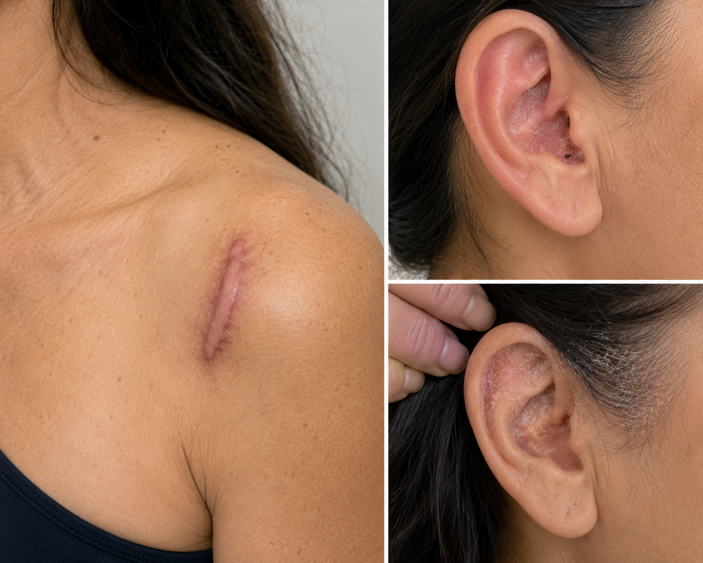
Recently, a 58-year-old woman visited our dermatology clinic with a raised scar on her left shoulder that developed after a fall approximately nine months earlier. During the same visit, she also sought treatment for persistent flaky, irritated ears that had stopped responding to her previous eczema medication.
After a comprehensive evaluation, our board-certified dermatologist diagnosed a hypertrophic scar and seborrheic dermatitis, creating a personalized treatment plan to address both conditions.
If you're looking for expert scar treatment or help with chronic flaky ears in Houston or Katy, Texas, here's what you should know.
The Patient's Story
Nine months after a fall, this patient continued to have a thickened scar on the front of her left shoulder.
Although the scar was stable, she wanted to know whether it could be softened and made less noticeable.
She also reported:
Burning skin inside the ears
Flaking
Pain
Occasional bleeding
She had previously been using mometasone cream, but it was no longer providing adequate relief.
Following examination with dermoscopy, two separate diagnoses were made:
Hypertrophic scar on the left shoulder
Seborrheic dermatitis involving the ears
What Is a Hypertrophic Scar?
A hypertrophic scar develops when the body produces too much collagen during wound healing.
Unlike a normal scar, it becomes:
Raised
Thickened
Firm
Sometimes itchy
Occasionally painful or tender
Unlike keloids, hypertrophic scars remain confined to the original area of injury and do not grow beyond the wound's borders.
These scars commonly develop after:
Falls
Surgical incisions
Burns
Cuts
Traumatic injuries
Can a Hypertrophic Scar Be Removed Completely?
This is one of the most common questions dermatologists hear.
Unfortunately, no treatment can completely erase a scar.
However, several therapies can significantly improve:
Thickness
Firmness
Itching
Tenderness
Overall appearance
The goal is to make the scar flatter, softer, and less symptomatic.
Intralesional Kenalog (ILK): A Proven Scar Treatment
After discussing available options, the patient chose to proceed with an intralesional Kenalog (ILK) injection.
Kenalog is a corticosteroid injected directly into the scar to help:
Flatten thickened scar tissue
Reduce excess collagen production
Improve itching and discomfort
Soften the scar over time
For this visit:
One hypertrophic scar was treated.
Kenalog 20 mg/mL was injected.
A total of 0.1 mL was administered.
Before treatment, the potential risk of skin thinning (atrophy) was reviewed, as this is a known but generally uncommon side effect when injections are performed appropriately.
Many patients require multiple treatment sessions to achieve the best cosmetic outcome.
Silicone Scar Sheets: An Important At-Home Treatment
In addition to the steroid injection, the patient was advised to use silicone scar sheets.
Silicone therapy is one of the most widely recommended non-invasive treatments for hypertrophic scars and may help:
Flatten raised scars
Improve scar texture
Decrease redness
Reduce itching
When used consistently over several months, silicone sheets can enhance the results of in-office treatments.
Why Were the Flaky Ears Diagnosed as Seborrheic Dermatitis?
The patient's ear symptoms had initially been treated as eczema, but the examination was more consistent with seborrheic dermatitis, a chronic inflammatory skin condition.
Seborrheic dermatitis commonly affects areas rich in oil glands, including:
The scalp
The eyebrows
Around the nose
Behind and inside the ears
Symptoms often include:
Flaking
Redness
Burning
Itching
Crusting
The condition tends to flare periodically and requires ongoing management.
Why Was Her Medication Changed?
Because mometasone cream was no longer adequately controlling her symptoms, her dermatologist recommended switching to:
Fluocinonide 0.05% topical solution
The liquid formulation is often easier to apply around and inside the ears than a cream and can provide better penetration in areas with hair or narrow skin folds.
She was instructed to:
Apply it twice daily during flares
Limit use to 14 days per month
Contact the office if she preferred a cream formulation instead
Managing Seborrheic Dermatitis at Home
In addition to prescription medication, patients with seborrheic dermatitis often benefit from:
Regular use of fragrance-free moisturizers
Medicated shampoos containing zinc pyrithione, selenium sulfide, or coal tar (when appropriate)
Avoiding excessive scratching
Managing stress, which may trigger flare-ups
Although seborrheic dermatitis is chronic, consistent treatment can greatly reduce symptoms and improve quality of life.
Dermatology Care for Scars and Seborrheic Dermatitis in Houston and Katy, Texas
Whether you're dealing with a raised scar after an injury or chronic flaky, irritated ears, a proper diagnosis can lead to more effective treatment. From intralesional steroid injections to customized therapies for seborrheic dermatitis, early intervention can improve both comfort and cosmetic outcomes.
At Village Dermatology, our board-certified dermatologists provide expert care for hypertrophic scars, keloids, seborrheic dermatitis, eczema, and other chronic skin conditions. We proudly serve patients throughout Houston and Katy, Texas with personalized treatment plans designed to help them look and feel their best.
If you have a raised scar or persistent flaky ears that aren't improving, schedule an appointment with Village Dermatology for a comprehensive evaluation.
Pimples on the Buttocks and Groin? It May Not Be Acne: Understanding Folliculitis
A 44-year-old woman developed persistent bumps on the buttocks and groin that were initially treated as a fungal infection. Learn how dermatologists diagnosed folliculitis, why the treatment changed, and how Village Dermatology helps patients in Houston and Katy, Texas.
Many adults assume that bumps on the buttocks or groin are simply acne. However, these areas are commonly affected by folliculitis, a condition that can closely resemble acne but requires a different treatment approach.
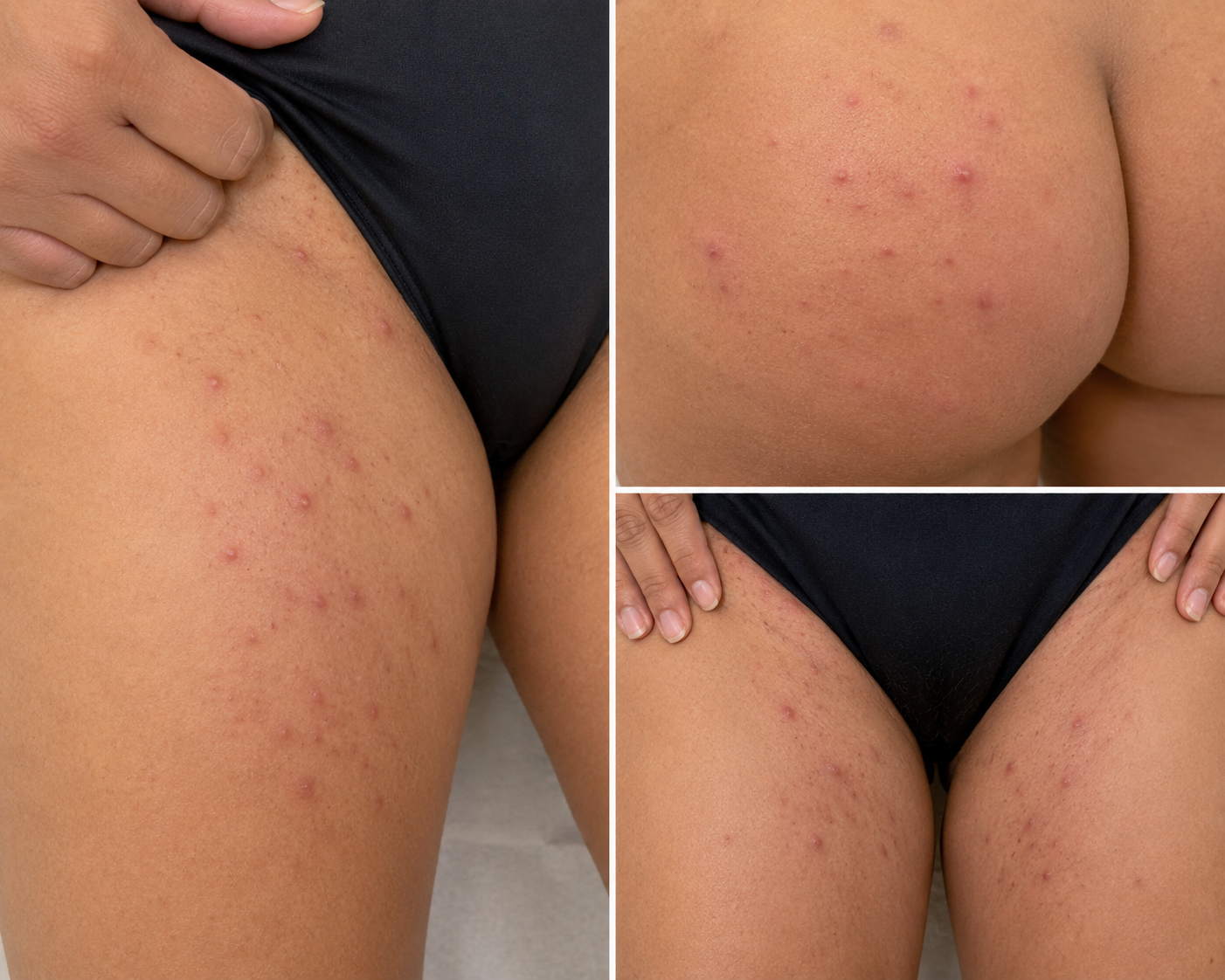
Recently, a 44-year-old woman visited our dermatology clinic after struggling with painful, inflamed bumps in her groin and buttock area for six months. She had already been treated with an antifungal medication without improvement, raising an important question:
If it's not a fungal infection, what is causing these bumps?
After a comprehensive skin examination, our board-certified dermatologist diagnosed folliculitis and developed a targeted treatment plan designed to eliminate the infection and reduce future flare-ups.
If you're searching for answers about recurring bumps in sensitive areas, here's what you should know.
The Patient's Story
This patient came to our clinic concerned about persistent "acne" affecting the:
Buttocks
Groin
For approximately six months, she had experienced:
Red inflamed bumps
Pimples
Recurrent flare-ups
She had previously been prescribed an oral antifungal medication (terbinafine) and instructed to use antifungal powder and Epsom salt baths. Despite these treatments, the bumps persisted.
A detailed examination, including dermoscopy, revealed follicular-based pustules, findings much more consistent with folliculitis than acne or a fungal infection.
Because of this, her treatment plan was changed.
What Is Folliculitis?
Folliculitis is inflammation or infection of the hair follicles.
It often appears as:
Small red bumps
White-headed pimples
Tender pustules
Itchy or painful lesions
Unlike traditional acne, folliculitis commonly develops in areas exposed to:
Friction
Sweat
Moisture
Tight-fitting clothing
Shaving
The buttocks and groin are among the most frequently affected areas.
Why Wasn't This a Fungal Infection?
Although fungal infections can occasionally mimic folliculitis, several features suggested this patient's bumps were instead caused by inflamed hair follicles.
Because her lesions appeared follicular in nature and had not improved with antifungal therapy, continuing antifungal powders was unlikely to provide benefit.
Instead, treatment was directed toward the more likely diagnosis of bacterial folliculitis.
Why Does Folliculitis Develop?
Folliculitis may occur when hair follicles become irritated or infected.
Common contributing factors include:
Heat and humidity
Excessive sweating
Tight athletic clothing
Prolonged sitting
Friction from exercise
Shaving or waxing
Bacterial overgrowth on the skin
Sometimes multiple factors work together, leading to recurrent flare-ups.
Treatment Plan
Because the lesions were consistent with folliculitis, the treatment plan focused on reducing bacteria and inflammation.
Oral Doxycycline
The patient was prescribed:
Doxycycline 100 mg twice daily for four weeks.
Doxycycline helps reduce both bacterial growth and inflammation, making it an effective option for moderate folliculitis.
Antibacterial Cleansers
Daily cleansing was recommended using antibacterial washes, including:
Hibiclens® (chlorhexidine)
CLn® SportWash (alternating regimen)
These cleansers help decrease bacterial colonization on the skin and may reduce future outbreaks.
Patients may also benefit from a benzoyl peroxide wash, which can help decrease bacteria within hair follicles.
Stop Unnecessary Antifungal Treatment
Because the examination favored folliculitis rather than a fungal infection, the patient was advised that ongoing antifungal powders were no longer necessary.
Avoiding unnecessary medications helps simplify treatment and focuses therapy on the underlying cause.
Doxycycline Safety Tips
Patients taking doxycycline should follow several important precautions.
Our dermatologist reviewed:
Take the medication with food and plenty of water.
Avoid lying down immediately after taking each dose.
Increase sun protection because doxycycline can increase sensitivity to sunlight.
Wear SPF 30+ sunscreen, protective clothing, and sunglasses when outdoors.
Contact your healthcare provider if severe headaches, vision changes, hearing changes, or unusual bruising occur.
Avoid pregnancy while taking doxycycline due to potential risks to a developing baby.
Understanding these precautions helps maximize treatment effectiveness while minimizing side effects.
Preventing Future Folliculitis
Many patients can reduce recurrence by making a few simple lifestyle changes.
Helpful habits include:
Showering promptly after exercise
Wearing loose, breathable clothing
Changing out of sweaty workout clothes quickly
Avoiding excessive friction in affected areas
Using antibacterial cleansers as directed
Keeping the skin clean and dry
For patients with frequent flare-ups, an individualized prevention plan can make a significant difference.
When Should You See a Dermatologist?
While mild folliculitis may resolve on its own, medical evaluation is recommended if you experience:
Recurrent outbreaks
Painful or draining bumps
Lesions lasting several weeks
Scarring
Failure to improve with over-the-counter products
Uncertainty about whether the condition is acne, folliculitis, hidradenitis suppurativa, or a fungal infection
An accurate diagnosis is essential because these conditions often require very different treatments.
Folliculitis Treatment in Houston and Katy, Texas
Persistent bumps in the groin or buttocks can be frustrating and uncomfortable, but effective treatment begins with the correct diagnosis. Conditions that appear to be "acne" are often folliculitis or another inflammatory skin disorder that responds well to targeted medical therapy.
At Village Dermatology, our board-certified dermatologists diagnose and treat folliculitis, acne, hidradenitis suppurativa, fungal skin conditions, and other disorders affecting sensitive areas. We provide personalized treatment plans for patients throughout Houston and Katy, Texas, helping them achieve healthier skin and prevent recurrent flare-ups.
If you're struggling with recurring bumps on the buttocks, groin, or other areas of the body, schedule an appointment with Village Dermatology for a comprehensive evaluation.
Multiple Infantile Hemangiomas in a Baby: Why Did Our Dermatologist Recommend a Liver Ultrasound?
A 9-month-old infant presented with multiple infantile hemangiomas on the back, buttock, and toe. Learn why a liver ultrasound may be recommended, when treatment is needed, and how pediatric dermatologists in Houston and Katy, Texas evaluate these common vascular birthmarks.
Seeing bright red spots appear on your baby's skin can be alarming for any parent. While many of these marks are harmless birthmarks, some require a closer evaluation—especially when there are several of them.
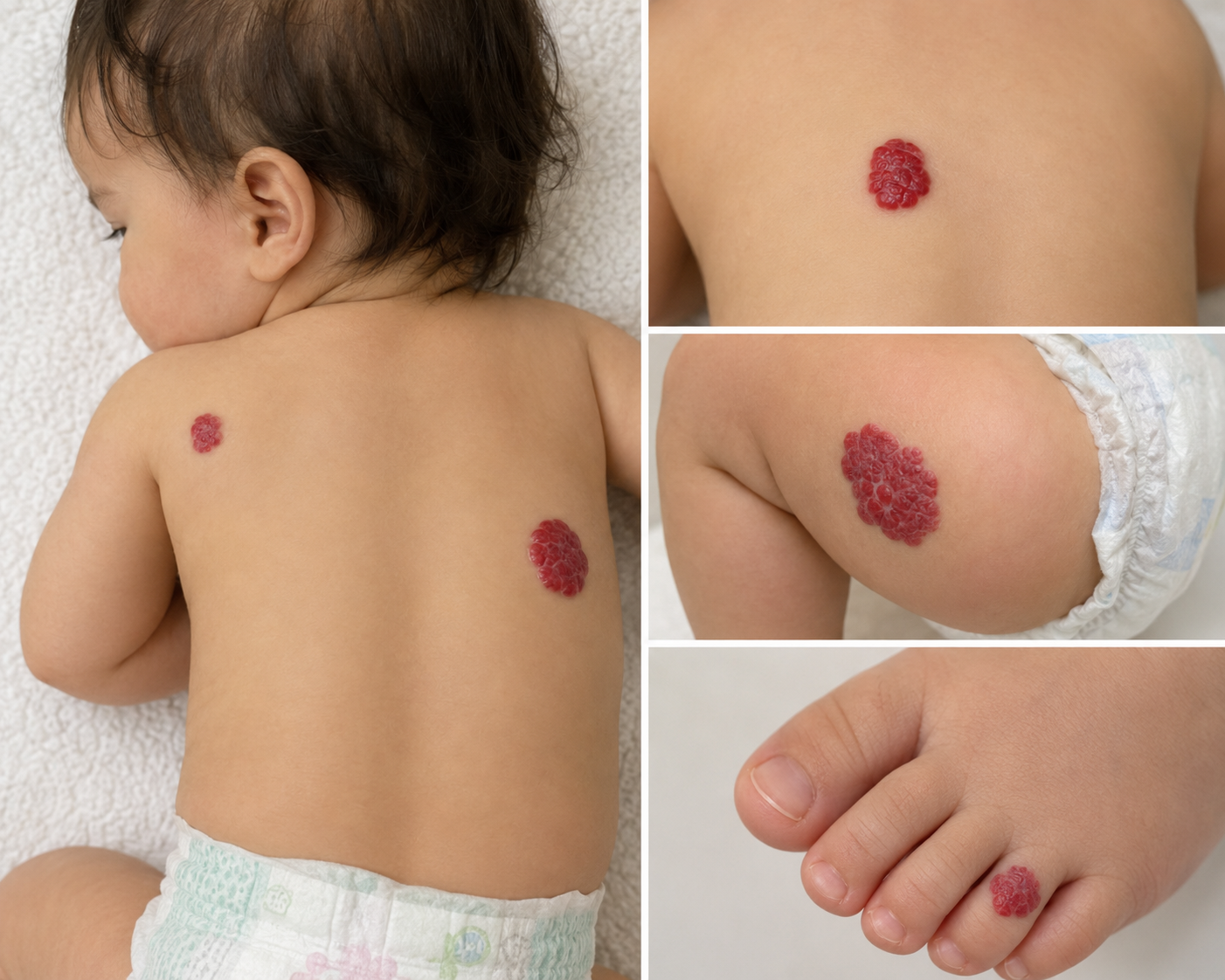
Recently, a 9-month-old female was brought to our dermatology clinic by her mother for evaluation of multiple red skin lesions located on her back, right buttock, and right fourth toe. Although the lesions were consistent with infantile hemangiomas, their number prompted an important discussion about additional screening to ensure there were no similar growths inside the body.
If your baby has multiple hemangiomas and you're searching for a pediatric dermatologist in Houston or Katy, Texas, here's what you should know.
The Patient's Story
A 9-month-old infant presented as a new patient for evaluation of several bright red vascular birthmarks.
Her mother was concerned because the lesions had appeared in multiple locations, including:
Left upper back
Right mid-back
Right buttock
Right fourth toe
A detailed skin examination, including dermoscopy, confirmed that these lesions were most consistent with infantile hemangiomas, the most common benign vascular tumors seen during infancy.
Because multiple hemangiomas were present, additional evaluation was recommended.
What Is an Infantile Hemangioma?
An infantile hemangioma is a benign collection of extra blood vessels that develops shortly after birth.
They often begin as:
Small red spots
Pink patches
Raised, bright-red bumps
Soft vascular growths
Most hemangiomas grow during the first several months of life before gradually shrinking over several years.
Many eventually fade significantly without requiring treatment.
Why Did Multiple Hemangiomas Lead to a Liver Ultrasound?
Most babies who have a single hemangioma require no additional testing.
However, when multiple infantile hemangiomas are present, dermatologists may recommend a liver ultrasound to look for possible hepatic hemangiomas.
Although uncommon, infants with several skin hemangiomas have a higher likelihood of also having hemangiomas within the liver.
Most hepatic hemangiomas cause no problems, but identifying them early allows appropriate monitoring and referral if needed.
For this patient, a liver ultrasound was ordered as a precautionary screening study.
What Happens If the Ultrasound Is Abnormal?
If liver imaging identifies multiple hepatic hemangiomas or another vascular abnormality, the child may be referred to a vascular anomalies clinic or pediatric specialist for further evaluation.
These multidisciplinary teams can determine whether observation alone is appropriate or whether treatment is recommended.
Fortunately, many infants never require intervention.
Do All Infantile Hemangiomas Need Treatment?
No.
Many infantile hemangiomas can simply be observed because they naturally go through two phases:
Growth Phase
During the first several months of life, the hemangioma enlarges.
Involution Phase
Over the following years, the blood vessels slowly shrink, and the lesion becomes flatter and lighter in color.
Because of this natural process, many children never require medication or surgery.
When Is Treatment Recommended?
Treatment may be considered when hemangiomas:
Grow rapidly
Bleed frequently
Become ulcerated or painful
Interfere with vision, breathing, feeding, or hearing
Are located in areas at high risk for scarring or functional problems
Cause significant cosmetic concerns
Early evaluation by a board-certified dermatologist helps determine whether treatment should begin during the growth phase.
What Treatment Options Were Discussed?
Several evidence-based options were reviewed with the patient's mother, including:
Topical Timolol
A beta-blocker medication that may be effective for certain small, superficial hemangiomas.
Oral Propranolol
The first-line treatment for many larger, deeper, rapidly growing, or high-risk infantile hemangiomas.
Oral propranolol has transformed the treatment of infantile hemangiomas and has an excellent safety profile when prescribed and monitored appropriately.
After discussing the risks, benefits, and expected outcomes, the family chose to defer medication while proceeding with the recommended liver ultrasound and observation.
Why Regular Follow-Up Matters
Even when treatment is not started immediately, routine follow-up allows your dermatologist to:
Monitor growth
Detect complications early
Determine whether treatment becomes necessary
Reassure families as lesions begin to naturally regress
Most infantile hemangiomas improve significantly with time, but careful monitoring ensures that babies who need treatment receive it at the optimal stage.
Pediatric Dermatology Care in Houston and Katy, Texas
Infantile hemangiomas are common, but every child deserves an individualized evaluation. When multiple hemangiomas are present, a thorough skin examination and appropriate screening can provide reassurance while identifying the rare infants who need additional care.
At Village Dermatology, our board-certified dermatologists provide expert evaluation and management of infantile hemangiomas, vascular birthmarks, and other pediatric skin conditions for families throughout Houston and Katy, Texas.
If your baby has a growing red birthmark or multiple vascular lesions, schedule an appointment with our team for a comprehensive evaluation and personalized treatment plan.
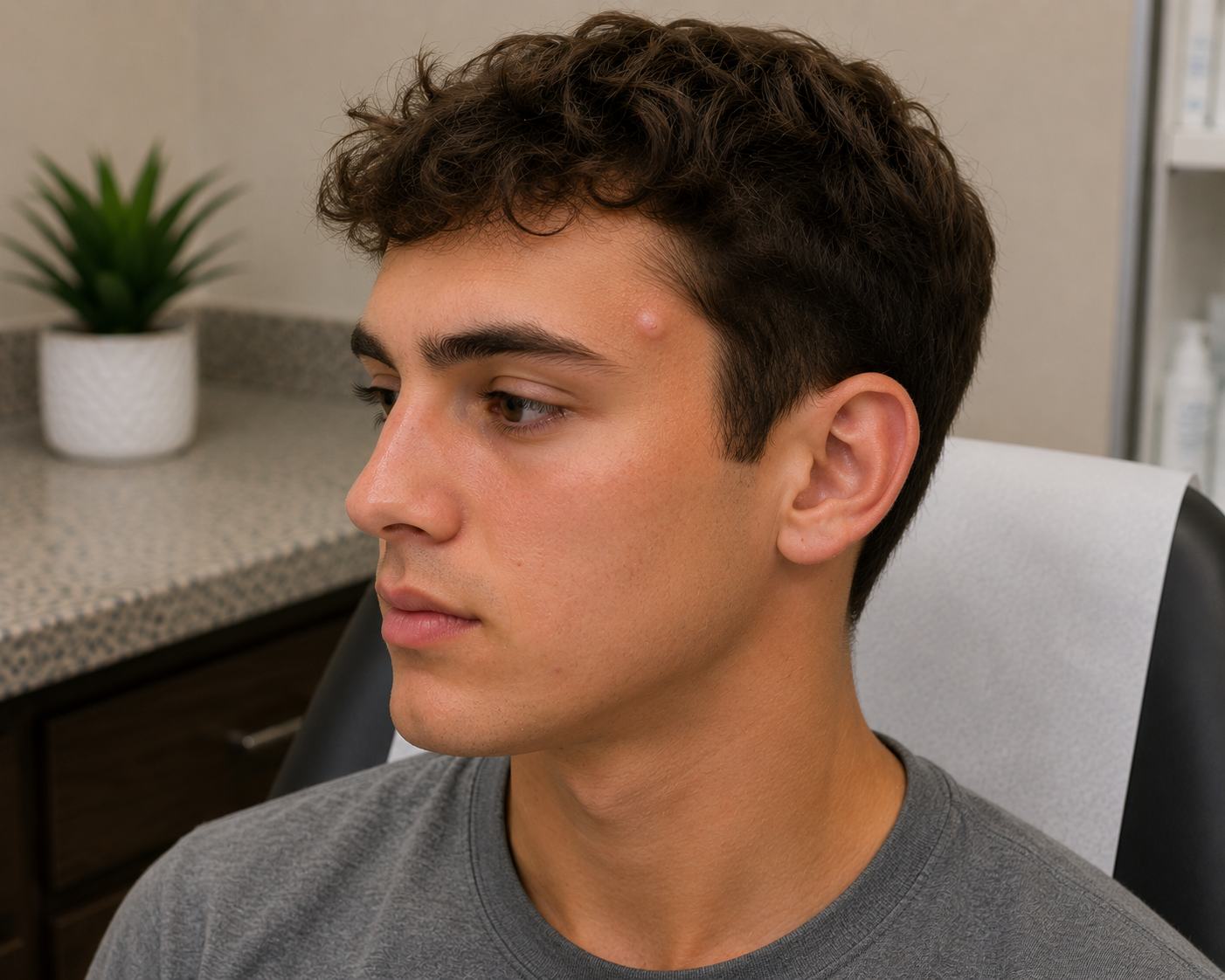
Why Am I Still Getting Pimples During Month 3 of Accutane? A Houston Dermatologist Explains
A 19-year-old patient continued improving during month three of Accutane despite experiencing a few new breakouts. Learn why occasional acne during isotretinoin treatment is normal, how monthly monitoring keeps treatment safe, and what to expect from acne care at Village Dermatology in Houston and Katy, Texas.
Acne can be frustrating enough, but it can feel even more discouraging when you're already taking Accutane (isotretinoin) and still notice a few new breakouts. Many patients expect their skin to become completely clear within the first couple of months, only to discover that occasional pimples can still appear.
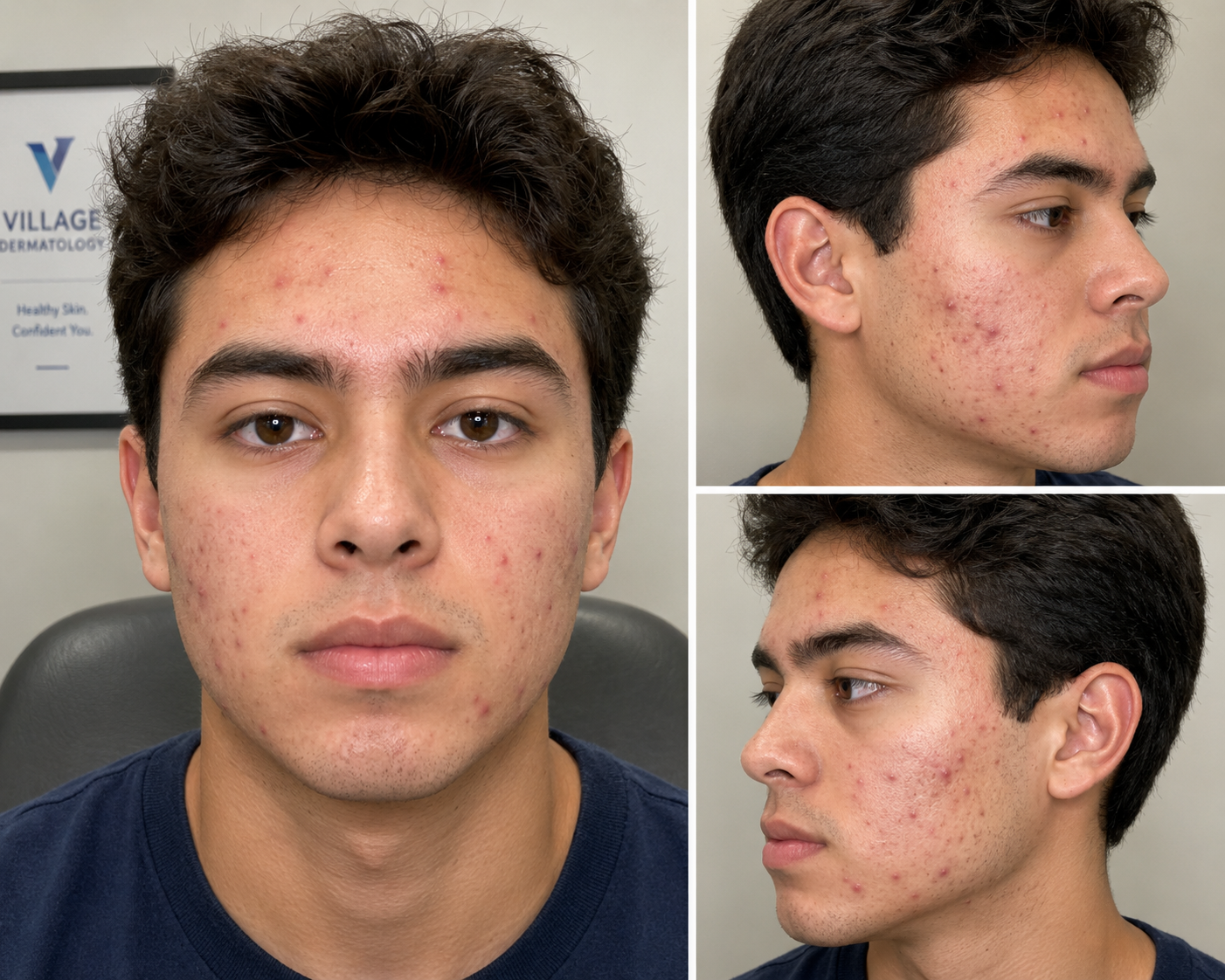
Recently, a 19-year-old male visited our dermatology clinic for his third month of isotretinoin treatment after beginning therapy for moderate inflammatory acne. Although his acne was improving and he was tolerating treatment well, he was concerned because he had developed a few new breakouts over the previous month.
Fortunately, this is a very common part of the Accutane journey.
If you're searching for an Accutane dermatologist in Houston or Katy, Texas, here's what you should know about treatment expectations and why patience often leads to the best long-term results.
The Patient's Story
This patient returned for his routine monthly isotretinoin follow-up after starting 80 mg daily (40 mg twice daily with a fatty meal).
During his visit, he reported:
Only manageable skin dryness
No serious medication side effects
A few new acne breakouts during the past month
Good overall tolerance of treatment
On examination, there were still:
Inflammatory papules
Pustules
Comedonal acne (blackheads and whiteheads)
primarily affecting the face.
Because he was responding appropriately and tolerating therapy well, his dermatologist recommended continuing isotretinoin at 80 mg daily while monitoring his progress.
Is It Normal to Break Out During Month Three?
Yes.
Many patients continue to develop occasional pimples during the first several months of isotretinoin therapy.
Although isotretinoin is the most effective medication available for severe acne, it does not eliminate acne overnight.
Instead, it gradually works by:
Shrinking oil glands
Dramatically reducing oil production
Preventing clogged pores
Reducing acne-causing bacteria
Decreasing inflammation deep within the skin
As these changes occur, the skin progressively becomes clearer over several months.
When Will My Skin Actually Clear?
Most patients begin noticing meaningful improvement after 2 to 3 months of treatment.
Significant improvement often occurs between:
Month 3
Month 4
Month 5
Many patients continue improving until the end of therapy.
For this reason, dermatologists emphasize that treatment success should be judged over the entire course, not month-to-month.
Why Was Treatment Continued Instead of Increased?
This patient's treatment protocol was already appropriate for his weight.
His dermatologist determined that:
Side effects were minimal
Acne was improving
Only a few new lesions remained
Treatment remained well tolerated
Because of this, continuing the current dose was the safest and most effective decision.
Sometimes maintaining a consistent dose provides better long-term results than making unnecessary adjustments.
Why Does Accutane Require Monthly Visits?
Isotretinoin is considered a high-risk medication, meaning patients require careful medical supervision throughout treatment.
Monthly follow-up visits allow your dermatologist to:
Monitor treatment progress
Evaluate side effects
Adjust medication if needed
Review skincare recommendations
Ensure medication safety
Order laboratory monitoring when appropriate
For this patient, a hepatic function panel and lipid panel were ordered as part of routine isotretinoin monitoring.
These tests help ensure the medication continues to be used safely.
Managing Dryness During Accutane
Dry skin remains the most common side effect of isotretinoin.
Fortunately, this patient's dryness was mild and manageable.
Patients can often improve comfort by using:
Gentle non-comedogenic cleansers
Oil-free moisturizers
Lip balm throughout the day
Broad-spectrum SPF 30+ sunscreen every morning
Maintaining a consistent skincare routine helps patients stay comfortable throughout treatment.
Important Accutane Safety Reminders
During every follow-up visit, patients receive ongoing education about isotretinoin safety.
Key reminders include:
Never share your medication.
Do not donate blood while taking isotretinoin.
Complete recommended laboratory monitoring.
Notify your dermatologist about any concerning side effects.
Report vision changes, severe headaches, mood changes, or significant muscle pain promptly.
Take isotretinoin with a fatty meal to improve absorption.
These precautions help maximize both safety and treatment effectiveness.
The Importance of Reaching the Target Cumulative Dose
One reason dermatologists encourage patients to stay the course is that isotretinoin works best when a patient reaches their target cumulative dose.
This patient had completed approximately three months of therapy and had accumulated 24 mg/kg, with a long-term treatment goal of approximately 200–220 mg/kg under his dermatologist's treatment protocol.
Reaching the recommended cumulative dose has been associated with a lower risk of acne recurrence after treatment is complete.
Acne Treatment in Houston and Katy, Texas
Acne affects far more than your skin—it can impact confidence, social interactions, and quality of life. Fortunately, isotretinoin remains one of the most effective treatments for severe, persistent, or treatment-resistant acne when prescribed and monitored by an experienced dermatologist.
At Village Dermatology, our board-certified dermatologists provide personalized acne treatment plans, including comprehensive isotretinoin monitoring, laboratory testing, and guidance to help patients achieve long-term clearance safely.
If you're struggling with stubborn acne or wondering whether Accutane is right for you, we're here to help.
Schedule an appointment at our Houston or Katy, Texas locations to discuss your options and develop a treatment plan tailored to your skin.
"I'm Two Months Into Accutane and I'm Still Getting Pimples—Is My Treatment Actually Working?"
A 29-year-old man developed significant hair shedding after losing 25 pounds. Learn how rapid weight loss can trigger telogen effluvium while revealing underlying male pattern hair loss, and discover effective treatment options at Village Dermatology in Katy and Houston, Texas.
Starting accutane® (isotretinoin) is often a life-changing decision for people who have struggled with severe acne for years. But many patients become discouraged when they continue to develop new pimples during the first few months of treatment.
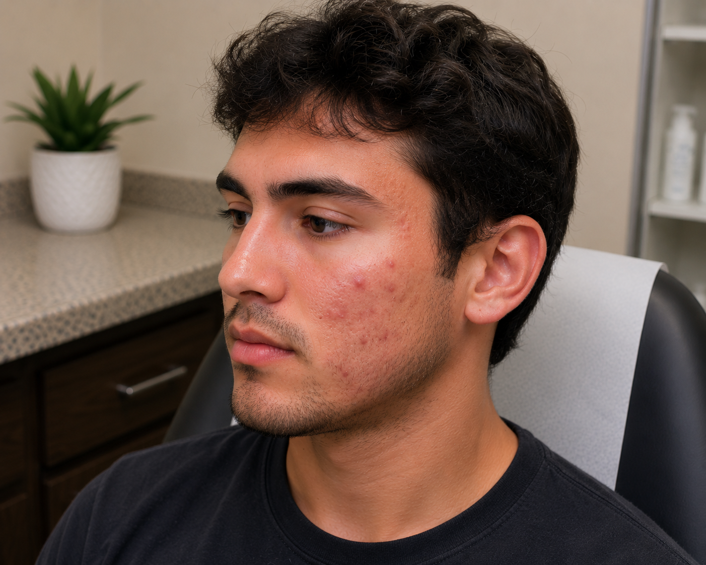
Recently, a 22-year-old man returned to our dermatology clinic in Houston for his second month of isotretinoin therapy. Before starting treatment, he had inflammatory acne affecting both cheeks along with acne scarring. Although he had noticed a few new breakouts over the past month, he was pleased to see that his skin was gradually improving overall.
After evaluating his progress, our board-certified dermatologist determined that his treatment was working as expected and recommended continuing his 100 mg daily isotretinoin regimen while performing routine laboratory monitoring.
If you're worried because you're still breaking out early in your Accutane journey, here's why that doesn't necessarily mean the medication isn't working.
Is It Normal to Break Out During the First Few Months of Accutane?
Yes.
One of the most common misconceptions about isotretinoin is that acne should disappear immediately after starting treatment.
In reality, many patients continue developing new pimples during the first 8 to 12 weeks. Some even experience a temporary flare before their skin begins clearing.
This happens because isotretinoin is gradually shrinking the oil glands beneath the skin while existing clogged pores continue working their way to the surface.
For most patients, meaningful improvement becomes much more noticeable after several months of consistent therapy.
How Does Isotretinoin Work?
Isotretinoin is considered the most effective medication available for severe acne because it targets every major cause of acne.
It works by:
Dramatically reducing oil production
Preventing clogged pores
Decreasing acne-causing bacteria
Reducing inflammation
Lowering the risk of future acne scarring
Unlike many topical medications, isotretinoin has the potential to produce long-term remission after a complete treatment course.
Why This Patient Stayed on the Same Dose
During this follow-up visit, the patient reported:
Only manageable dry skin (xerosis)
A few new pimples
Overall improvement in acne
Because he was tolerating treatment well without significant side effects, his dermatologist recommended continuing his current 100 mg daily dose.
Maintaining an effective dose helps patients steadily work toward their target cumulative dose while minimizing interruptions in treatment.
Why Does My Dermatologist Order Monthly Blood Tests?
Isotretinoin is a highly effective medication, but it requires careful monitoring throughout treatment.
Routine laboratory testing helps evaluate:
Liver function
Triglyceride levels
Overall medication safety
Most patients complete these blood tests once a month while taking isotretinoin.
Monitoring allows dermatologists to identify any significant changes early while ensuring patients can safely continue treatment.
Why Does the Total Cumulative Dose Matter?
Rather than simply treating acne until the skin clears, dermatologists often calculate a target cumulative dose based on body weight.
Many treatment plans aim for approximately 200–220 mg per kilogram over the course of therapy.
Reaching this cumulative exposure has been shown to reduce the likelihood that acne will return after treatment is completed.
This patient's treatment plan was designed using this evidence-based approach.
Managing Common Side Effects
Fortunately, this patient experienced only mild dryness, which is the most common side effect of isotretinoin.
Simple measures can make treatment much more comfortable, including:
Applying fragrance-free moisturizer daily
Using lip balm frequently
Washing with a gentle cleanser
Wearing broad-spectrum SPF 30+ sunscreen every morning
Avoiding harsh exfoliants and drying acne products
These supportive skincare habits allow many patients to remain comfortable throughout therapy.
Important Safety Reminders While Taking Accutane
Every follow-up visit includes important counseling to help patients use isotretinoin safely.
Patients are advised to:
Never share their medication.
Avoid donating blood while taking isotretinoin.
Report any unusual symptoms immediately.
Be cautious with nighttime driving if vision changes occur.
Delay elective cosmetic procedures or surgery for several months after completing treatment if advised by their dermatologist.
Following these recommendations helps reduce complications while maximizing treatment success.
Patience Leads to Clearer Skin
One of the most encouraging aspects of this visit was that, despite a few new pimples, the patient's acne was steadily improving.
This is exactly what dermatologists hope to see during the second month of treatment.
Consistency, regular follow-up appointments, laboratory monitoring, and patience are the keys to achieving long-term results.
Many patients who complete a full course of isotretinoin experience dramatic, long-lasting improvement and significantly lower rates of future acne breakouts.
Expert Accutane Treatment in Katy & Houston, Texas
If severe acne is affecting your confidence or leaving permanent scars, you don't have to manage it alone.
At Village Dermatology, our board-certified dermatologists provide comprehensive isotretinoin (Accutane®) treatment, including personalized dosing, iPLEDGE management, monthly laboratory monitoring, and ongoing support throughout your treatment journey.
Whether you're just beginning Accutane or seeking expert guidance after previous acne treatments have failed, our experienced team proudly serves patients throughout Houston, Katy, and surrounding Texas communities.
Schedule your consultation today and discover whether isotretinoin is the right solution for achieving clearer, healthier skin.
"I Lost 25 Pounds and Now My Hair Is Falling Out—Is the Weight Loss to Blame?"
A 22-year-old man continued to experience a few breakouts during his second month of Accutane while showing overall improvement. Learn why early breakouts are common, why monthly blood tests matter, and what to expect from isotretinoin treatment at Village Dermatology in Katy and Houston, Texas.
Losing weight can be a major accomplishment for your overall health—but for some people, it comes with an unexpected side effect: noticeable hair shedding.
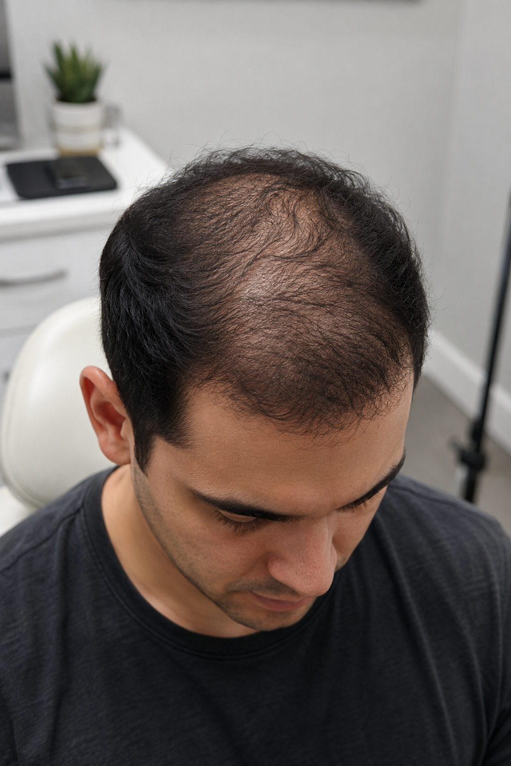
Recently, a 29-year-old man visited our dermatology clinic in Houston after experiencing three months of diffuse hair thinning across his scalp. He had become increasingly concerned by the amount of hair he was seeing in the shower and on his pillow. During his consultation, he shared that he had recently lost approximately 25 pounds, raising an important question:
Could rapid weight loss be causing his hair loss?
After a comprehensive scalp examination using dermoscopy, our board-certified dermatologist diagnosed telogen effluvium (TE) triggered by recent weight loss, along with early underlying androgenetic alopecia (male pattern hair loss). Understanding the difference between these two conditions was the key to creating an effective treatment plan.
If you've noticed increased shedding after dieting, illness, or significant weight changes, here's what you should know.
Can Weight Loss Really Cause Hair Loss?
Yes.
Rapid weight loss is one of the most common triggers for telogen effluvium, a temporary form of hair shedding.
When the body experiences significant physical stress—such as:
Rapid weight loss
Major surgery
High fever or illness
Emotional stress
Iron deficiency
Thyroid disorders
Certain medications
it shifts more hair follicles into the resting (telogen) phase of the hair growth cycle.
Approximately two to three months later, those resting hairs begin shedding all at once.
This timeline matched our patient's history almost perfectly.
What Is Telogen Effluvium?
Normally, about 85–90% of your hair follicles are actively growing while only a small percentage are resting.
With telogen effluvium, a larger number of follicles enter the resting phase simultaneously.
The result is:
Increased hair shedding
Hair found on pillows or clothing
More hair in the shower drain
Diffuse thinning rather than isolated bald patches
Importantly, the hair follicles remain alive, meaning regrowth is possible once the underlying trigger improves.
Why Was Hair Loss Most Noticeable on the Top of His Scalp?
Although this patient's shedding was caused by telogen effluvium, his examination also revealed early androgenetic alopecia (AGA).
Androgenetic alopecia is a genetic condition that gradually causes hair follicles to shrink over time.
It typically affects:
The frontal hairline
Temples
Crown (vertex)
Mid-scalp
When telogen effluvium occurs in someone with early male pattern baldness, the increased shedding often makes the genetic hair thinning appear much worse.
Many patients think they've suddenly developed permanent baldness when, in reality, two different conditions are occurring at the same time.
Diagnosing Two Types of Hair Loss
Using dermoscopy and a detailed scalp examination, the dermatologist identified:
Diffuse increased shedding consistent with telogen effluvium
Patterned thinning involving the frontal scalp consistent with androgenetic alopecia
Recognizing both diagnoses is important because each condition requires a slightly different treatment approach.
Why Oral Minoxidil Was Recommended
After discussing multiple treatment options, the patient elected to begin oral minoxidil 2.5 mg once daily.
Low-dose oral minoxidil has become an increasingly popular treatment for many forms of hair loss because it helps stimulate follicles back into the active growth phase.
It may benefit patients with:
Telogen effluvium lasting longer than expected
Male pattern hair loss
Female pattern hair loss
Combined hair loss conditions
Because oral minoxidil is a prescription medication, patients should be monitored by an experienced dermatologist.
Does Minoxidil Cause More Shedding at First?
One of the most important counseling points involved preparing the patient for a temporary increase in shedding.
During the first 8 to 10 weeks, some patients notice additional hair loss after starting minoxidil.
Although this can be alarming, it often represents older resting hairs being shed so healthier hairs can begin growing.
This temporary shedding is generally considered a normal part of treatment and does not mean the medication is making hair loss worse.
Other Hair Restoration Options
The patient also discussed several additional treatment options that can complement medical therapy, including:
Topical 5% minoxidil
Finasteride
Dutasteride
Platelet-Rich Plasma (PRP)
Alma TED™ hair restoration
Nutritional supplements
Low-Level Laser Therapy (LLLT)
Treatment recommendations depend on each patient's age, diagnosis, medical history, and long-term hair restoration goals.
Important Side Effects of Oral Minoxidil
Although most patients tolerate low-dose oral minoxidil well, it's important to understand potential side effects.
Patients should stop the medication and contact their dermatologist immediately if they experience:
Shortness of breath
Chest pain
Swelling of the ankles or feet
Rapid heartbeat
Dizziness
Low blood pressure
Fluid retention
Regular follow-up appointments allow dermatologists to monitor treatment progress and ensure the medication remains safe.
The Good News About Weight Loss-Related Hair Shedding
Unlike permanent forms of hair loss, telogen effluvium is usually self-limited.
Once the underlying trigger has resolved and the body has recovered, most patients gradually begin regrowing hair over several months.
However, when telogen effluvium uncovers underlying androgenetic alopecia, continued treatment may be recommended to preserve long-term hair density.
Early diagnosis gives patients the best opportunity to maintain and restore healthy hair.
Expert Hair Loss Treatment in Katy & Houston, Texas
Hair shedding after weight loss can be frightening, but you don't have to figure it out on your own.
At Village Dermatology, our board-certified dermatologists specialize in diagnosing every type of hair loss, including telogen effluvium, androgenetic alopecia, alopecia areata, and scarring alopecias. We offer personalized treatment plans using evidence-based therapies such as oral and topical minoxidil, finasteride, PRP, Alma TED™, nutritional guidance, and advanced scalp evaluations.
If you're noticing increased shedding, thinning, or changes in your hairline, schedule a consultation with our experienced team serving Houston, Katy, and surrounding Texas communities. Early treatment can make a significant difference in preserving your hair and restoring your confidence.
"I Have a Soft Lump on My Temple That's Been There for Years—Do I Need to Have It Removed?"
A 19-year-old man sought evaluation for a soft, painless lump on his temple. Learn how dermatologists diagnose epidermal inclusion cysts, lipomas, and dermoid cysts, and when surgical removal is recommended at Village Dermatology in Katy and Houston, Texas.
Finding a lump beneath your skin can be unsettling, especially when it's on your face. Even if it isn't painful, many people worry that it could be something serious or wonder whether it will continue to grow over time.
Recently, a 19-year-old man visited our dermatology clinic in Houston with a soft bump on his left temple that had been present for several years. Although the lesion wasn't causing pain or other symptoms, he wanted to know exactly what it was and whether it should be removed.
After a careful examination, our board-certified dermatologist determined that the mass was most consistent with either an epidermal inclusion cyst (EIC) or a lipoma, while also considering the possibility of a dermoid cyst. Because the lesion measured approximately 1.7 × 1.2 cm and was located on the face, surgical excision was recommended both to establish a definitive diagnosis and remove the growth.
If you've discovered a painless lump on your face or scalp, here's what you should know.
What Is an Epidermal Inclusion Cyst?
An epidermal inclusion cyst (EIC) is one of the most common benign skin growths seen by dermatologists.
These cysts develop when skin cells become trapped beneath the surface instead of shedding normally. The trapped cells continue producing keratin, a natural skin protein, causing the cyst to slowly enlarge over time.
Most epidermal inclusion cysts are:
Soft or slightly firm
Round or oval
Mobile beneath the skin
Slow growing
Usually painless
Although harmless, they can become inflamed or infected if they rupture.
Could It Be a Lipoma?
Another possibility discussed during this patient's visit was a lipoma.
Lipomas are benign tumors made up of normal fat cells.
Compared with cysts, lipomas are often:
Very soft
Easily movable
Located deeper beneath the skin
Slow growing
Usually painless
Because both cysts and lipomas can feel similar during an examination, distinguishing between them sometimes requires surgical removal and laboratory analysis.
What Is a Dermoid Cyst?
Because the lesion was located near the temple, a dermoid cyst was also included in the differential diagnosis.
Dermoid cysts are congenital growths that develop before birth and may contain skin structures such as:
Hair follicles
Oil glands
Keratin
Although many dermoid cysts remain stable for years, they are often removed to confirm the diagnosis and prevent gradual enlargement.
How Dermatologists Evaluate Facial Lumps
During this patient's examination, the dermatologist carefully assessed several characteristics of the lesion.
The bump was:
Soft
Freely mobile beneath the skin
Well-defined
Approximately 1.7 × 1.2 centimeters
Located on the left temple
Not inflamed or tender
A dermatoscope was also used to closely evaluate the overlying skin and help rule out other skin lesions.
These findings favored a benign diagnosis rather than skin cancer.
Do All Cysts Need to Be Removed?
Not necessarily.
Many epidermal inclusion cysts can simply be observed if they are:
Small
Not painful
Not infected
Not rapidly enlarging
Not cosmetically bothersome
However, removal is often recommended when a cyst:
Continues growing
Frequently becomes inflamed
Ruptures repeatedly
Causes discomfort
Is located in a cosmetically sensitive area such as the face
For this patient, elective surgical excision was recommended because of the lesion's size and facial location.
What Does Surgical Excision Involve?
Complete surgical excision is considered the most effective treatment for epidermal inclusion cysts and many lipomas.
During the procedure:
Local anesthetic is used to numb the area.
A small incision is made over the lesion.
The entire cyst or fatty tumor is carefully removed.
The incision is closed with sutures to promote optimal healing.
Completely removing the cyst wall significantly reduces the chance of recurrence.
Will There Be a Scar?
One of the most important parts of the discussion involved balancing the benefits of removing the lesion with the possibility of a scar.
Our dermatologist explained that while surgical excision removes the bump, it replaces it with a carefully planned surgical scar.
Fortunately, facial skin generally heals very well, and dermatologic surgeons use meticulous techniques to minimize visible scarring whenever possible.
For many patients, replacing a noticeable lump with a thin, well-healed scar provides a significant cosmetic improvement.
When Should You Seek Medical Attention?
Although most cysts are harmless, patients should schedule an evaluation if a lump:
Grows rapidly
Becomes painful
Turns red or warm
Begins draining material
Repeatedly becomes inflamed
Changes in appearance
Early evaluation allows dermatologists to determine whether observation or removal is the best option.
Expert Cyst Evaluation and Removal in Katy & Houston, Texas
If you've noticed a lump beneath your skin, don't ignore it or try to squeeze it at home.
At Village Dermatology, our board-certified dermatologists diagnose and treat epidermal inclusion cysts, lipomas, dermoid cysts, and other benign skin growths using advanced diagnostic techniques and precise surgical procedures designed to achieve excellent cosmetic outcomes.
Whether your concern is located on the face, scalp, neck, or body, our experienced team proudly serves patients throughout Houston, Katy, and surrounding Texas communities.
Schedule your consultation today to learn whether your skin growth can simply be monitored or whether removal is the best option for your long-term health and peace of mind.

"My Toddler Has Little Bumps That Keep Spreading—Are They Molluscum Contagiosum?"
A 3-year-old girl developed spreading molluscum contagiosum involving the trunk, thighs, and abdomen. Learn how dermatologists diagnose and safely treat molluscum with Cantharone® Plus at Village Dermatology in Katy and Houston, Texas.
It can be alarming for parents to notice new bumps appearing on their child's skin—especially when they seem to spread from one area of the body to another. Many parents initially think the bumps are insect bites, warts, or a rash, only to discover they're caused by a common childhood viral infection.
Recently, a 3-year-old girl was brought to our dermatology clinic in Houston after her mother noticed multiple bumps that had been spreading over the previous nine months. The lesions had become more numerous and occasionally itchy, involving the abdomen, thighs, chest, and genital area.
After a thorough skin examination, our board-certified dermatologist diagnosed molluscum contagiosum, a very common viral skin infection in children. Because several lesions were inflamed and continuing to spread, treatment with Cantharone® Plus (cantharidin) was recommended.
If your child has small, shiny bumps that won't go away, here's what you should know.
What Is Molluscum Contagiosum?
Molluscum contagiosum is a benign viral skin infection caused by a member of the poxvirus family.
It is extremely common in young children and usually appears as:
Small pink or flesh-colored bumps
Smooth, shiny surface
A tiny central indentation (called an umbilication or "dell")
Firm, dome-shaped appearance
Although the bumps are generally painless, they can become itchy, inflamed, or irritated as the immune system begins clearing the virus.
Why Do Molluscum Bumps Spread?
The virus spreads through direct skin-to-skin contact and by touching contaminated objects.
Children commonly spread molluscum by:
Scratching the bumps
Sharing towels or washcloths
Close contact with siblings
Playing with other children
Swimming and other activities involving shared surfaces
As the child scratches, the virus can spread to nearby healthy skin, causing new bumps to appear over time.
What Did the Dermatologist Find?
During this patient's examination, multiple classic molluscum lesions were identified on the:
Abdomen around the belly button
Suprapubic area
Chest (sternum)
Thighs
Trunk
Genital region
The lesions appeared as pink, shiny, dome-shaped papules with a central dimple, which are characteristic of molluscum contagiosum.
Several lesions had become inflamed and were enlarging, making treatment medically appropriate.
How Is Molluscum Treated?
Although molluscum often resolves on its own over many months, treatment may be recommended when lesions are:
Continuing to spread
Inflamed
Itchy
Located in sensitive areas
Causing discomfort
Increasing in number
Several treatment options are available, including:
Cantharidin (Cantharone®)
Cryotherapy
Curettage
Tape stripping
Observation in selected cases
The best treatment depends on the child's age, the number of lesions, and their location.
What Is Cantharidin?
For this patient, Cantharone® Plus was applied to 12 molluscum lesions.
Cantharidin is a topical medication that causes a controlled blister to form beneath the treated lesion. As the blister heals, the infected skin separates from the healthy skin, allowing the body to eliminate the virus.
Because the medication is applied in the dermatology office, treatment is quick and well tolerated by most children.
What Should Parents Do After Treatment?
Proper aftercare helps ensure the best results.
Parents were instructed to:
Leave the medication on for approximately 4 hours
Wash the treated areas thoroughly with soap and water afterward
Expect temporary redness or blistering
Avoid scratching the treated areas
These reactions are expected and usually indicate that the medication is working.
Why Did the Dermatologist Also Discuss Eczema?
Many children with molluscum develop molluscum dermatitis, a type of eczema that forms around the bumps.
This occurs because the immune system reacts to the viral infection.
If itchy eczema develops after treatment, the dermatologist recommended using triamcinolone as directed to calm the inflammation.
In addition, gentle skin care remains an important part of treatment.
Parents were advised to:
Bathe children in lukewarm water.
Use mild, fragrance-free cleansers.
Apply moisturizers immediately after bathing.
Moisturize at least two to three times daily.
Avoid scented detergents and fabric softeners.
Keep fingernails short to reduce scratching and further spread.
Will Molluscum Go Away?
Yes.
Even without treatment, molluscum contagiosum eventually resolves as the immune system recognizes and eliminates the virus.
However, this process may take several months to over a year, and during that time the infection can spread to additional areas of the skin or to close contacts.
Treatment often helps reduce the number of lesions, shorten the duration of infection, and decrease transmission to siblings or classmates.
When Should Parents Contact Their Dermatologist?
Parents should schedule a follow-up visit if:
New lesions continue appearing rapidly.
The bumps become significantly inflamed.
A widespread itchy rash develops.
Signs of infection such as pus, warmth, or severe pain occur.
The lesions fail to improve after treatment.
Follow-up appointments allow additional lesions to be treated if needed.
Expert Molluscum Treatment in Katy & Houston, Texas
If your child has bumps that continue spreading or won't go away, an accurate diagnosis is the first step toward effective treatment.
At Village Dermatology, our board-certified dermatologists diagnose and treat molluscum contagiosum, eczema, warts, viral rashes, and other common childhood skin conditions using safe, evidence-based therapies designed specifically for pediatric patients.
Whether your child needs reassurance, observation, or treatment with cantharidin, our experienced team proudly serves families throughout Houston, Katy, and surrounding Texas communities.
Schedule an appointment today to help your child get relief and prevent molluscum from continuing to spread.

"I Thought I Had Eczema—Why Is My Dermatologist Treating Me for Scabies Instead?"
A 43-year-old woman developed a persistent rash that didn't improve with steroid cream. Learn how scabies can mimic eczema, when dermatologists suspect a mite infestation, and what treatment involves at Village Dermatology in Katy and Houston, Texas.
An itchy rash can be frustrating, especially when it doesn't improve with prescription steroid creams. While many people assume that red, irritated patches are simply eczema, several skin conditions can look remarkably similar—including one that is highly contagious.
Recently, a 43-year-old woman visited our dermatology clinic in Houston after developing a persistent rash on her arms and trunk that had been present for about a month. She had already tried triamcinolone cream, but the rash continued to appear. She had no history of eczema or other chronic skin conditions and had even stopped taking a magnesium supplement to see if it was causing the rash—but nothing changed.
After carefully examining the affected areas, our board-certified dermatologist considered two leading possibilities: eczema (dermatitis) and scabies. Because the appearance of the rash and the patient's history raised concern for scabies, treatment was started promptly while educating the patient about preventing spread to others.
If you've been treating what you thought was eczema without improvement, here's why your dermatologist may consider scabies as another possible diagnosis.
Why Can Scabies Look Like Eczema?
Scabies is caused by tiny microscopic mites (Sarcoptes scabiei) that burrow into the outer layer of the skin.
The body's immune reaction to these mites produces symptoms that often resemble eczema, including:
Red patches
Small bumps
Itching
Irritated skin
Scratch marks
Because these symptoms overlap with many inflammatory skin conditions, scabies is frequently mistaken for eczema during its early stages.
What Is Dermatitis?
Dermatitis is a general term that describes inflammation of the skin.
It can develop for many reasons, including:
Allergic reactions
Irritating chemicals
Dry skin
Environmental triggers
Immune-related skin conditions
Dermatitis often causes:
Redness
Dryness
Scaling
Itching
Inflamed patches
Topical corticosteroids such as triamcinolone are commonly used to treat eczema, but if the underlying problem is actually scabies, steroid creams alone won't eliminate the mites.
Clues That Point Toward Scabies
During this patient's evaluation, several factors prompted consideration of scabies.
Her rash:
Appeared on the arms and trunk
Had persisted for about one month
Had not responded to topical steroid therapy
Occurred without a previous history of eczema
While additional testing is sometimes performed, dermatologists may begin treatment when clinical suspicion is high because delaying therapy can allow the infestation to spread to household members.
How Is Scabies Treated?
Scabies treatment focuses on eliminating both the mites and newly hatched mites.
This patient's treatment plan included two prescription medications:
Permethrin 5% Cream
Permethrin is considered one of the first-line treatments for scabies.
Patients are instructed to:
Apply the cream from the neck down to the feet
Leave it on overnight for approximately 8 hours
Wash it off the following morning
Repeat the treatment one week later
The second treatment helps eliminate mites that hatch after the first application.
Oral Ivermectin
Because scabies can sometimes be extensive or difficult to eradicate, oral ivermectin (Stromectol®) was also prescribed.
The medication is typically taken:
On an empty stomach
With a full glass of water
As a single dose
Repeated one week later
Using both treatments together may improve treatment success in appropriate patients.
Preventing Reinfestation
Treating the skin alone isn't enough.
Scabies mites can survive away from the body for a short period, making environmental cleaning an important part of treatment.
Patients are generally advised to:
Wash recently worn clothing, towels, and bedding in hot water.
Dry fabrics on the highest heat setting.
Seal unwashable items in a plastic bag for at least 72 hours.
Vacuum upholstered furniture if appropriate.
Just as importantly, household members and close physical contacts should often be treated at the same time, even if they are not yet experiencing symptoms. This helps prevent the infestation from cycling back and forth between family members.
What Side Effects Can Occur?
Most patients tolerate treatment very well.
Possible side effects of ivermectin include:
Nausea
Diarrhea
Dizziness
Mild itching
Temporary swelling of lymph nodes or extremities
Patients should contact their dermatologist if they experience concerning symptoms or have questions about treatment.
When Should You Return to Your Dermatologist?
Even after successful treatment, itching may continue for several weeks because the immune system is still reacting to dead mites.
However, patients should schedule a follow-up evaluation if:
New rashes continue appearing.
Symptoms worsen after treatment.
Household members continue developing symptoms.
The rash fails to improve after several weeks.
In some cases, additional evaluation may reveal another diagnosis such as eczema, allergic dermatitis, or another inflammatory skin disorder.
Expert Rash Diagnosis in Katy & Houston, Texas
Not every itchy rash is eczema—and not every rash is caused by an allergy.
At Village Dermatology, our board-certified dermatologists specialize in diagnosing difficult skin rashes, including eczema, allergic contact dermatitis, scabies, psoriasis, fungal infections, and other inflammatory skin conditions. Through careful examination and individualized treatment plans, we help patients find answers and lasting relief.
If you've been dealing with a persistent rash that isn't responding to treatment, schedule an appointment with our experienced dermatology team serving Houston, Katy, and surrounding Texas communities. Early diagnosis can make all the difference in getting your skin healthy again.
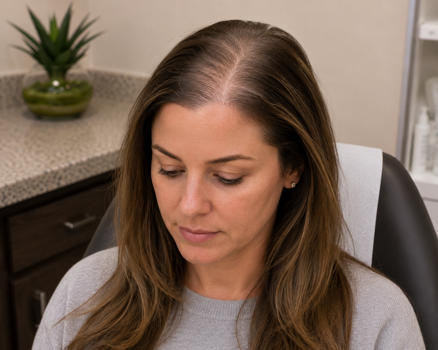
"I've Been Using Rogaine for Years and My Hair Is Still Thinning—What Are My Next Options?"
A 39-year-old woman with female pattern hair loss learned about today's most advanced treatment options beyond topical minoxidil, including oral minoxidil, PRP, Alma TED™, and regenerative hair restoration therapies at Village Dermatology in Katy and Houston, Texas.
For many women, noticing a widening part or increasing scalp visibility can be emotionally devastating. Even after faithfully using over-the-counter treatments like Rogaine®, some patients find that their hair continues to thin over time.
Recently, a 39-year-old woman returned to our dermatology clinic for follow-up of female pattern hair loss, also known as androgenetic alopecia (AGA). She had previously been using topical minoxidil (Rogaine®) and vitamin D supplementation but continued to notice gradual thinning over the top of her scalp.
During her visit, our board-certified dermatologist reviewed today's expanding list of medical and regenerative hair restoration treatments, helping her understand that while female pattern hair loss is chronic, there are now more effective treatment options than ever before.
If you've been wondering why your hair continues to thin despite using Rogaine®, here's what you should know.
What Is Female Pattern Hair Loss?
Female pattern hair loss is the most common cause of chronic hair thinning in women.
Unlike men, women usually don't develop complete bald spots. Instead, they experience:
A widening center part
Diffuse thinning across the crown
Decreased hair density
Increased scalp visibility
Gradual reduction in hair volume
The condition develops because genetically susceptible hair follicles slowly become smaller over time, producing finer, shorter hairs with each growth cycle.
Without treatment, the process usually progresses gradually over many years.
Why Didn't Rogaine Completely Stop My Hair Loss?
Topical minoxidil (Rogaine®) remains one of the first-line treatments for female pattern hair loss.
However, it's important to understand that:
It works only while you're using it.
Results typically take at least six months.
It slows progression more than it completely reverses hair loss.
Some patients eventually require additional therapies.
This patient had previously been instructed to use 5% minoxidil foam consistently because stopping treatment usually results in gradual loss of any regrown hair.
Newer Treatment Options Beyond Topical Minoxidil
During her follow-up appointment, several advanced treatment options were discussed.
Oral Minoxidil
Low-dose oral minoxidil has become an increasingly popular treatment for patients who want an alternative to daily topical applications.
By stimulating hair follicles internally, oral minoxidil may improve:
Hair density
Hair thickness
Overall scalp coverage
Potential side effects include:
Lightheadedness
Lower blood pressure
Swelling of the ankles
Increased body hair growth
Patients are carefully monitored throughout treatment.
PRP Hair Restoration
Platelet-Rich Plasma (PRP) therapy uses a patient's own blood to concentrate growth factors that are then injected into the scalp.
PRP may:
Stimulate dormant follicles
Reduce ongoing shedding
Increase hair thickness
Complement medical therapy
Many treatment plans include:
Four monthly sessions
Maintenance treatments every six months
Because PRP uses your own platelets, recovery is minimal, and patients can usually return to normal activities the same day.
Alma TED™ Hair Restoration
Another innovative option discussed was Alma TED™, a completely non-invasive hair restoration treatment.
Unlike injections, Alma TED delivers a specialized plant-based hair growth serum into the scalp using ultrasound and air pressure technology.
Benefits include:
No needles
No downtime
Comfortable treatment sessions
Support for healthier hair follicles
Can be combined with PRP or medications
Many patients appreciate Alma TED as an alternative if they prefer to avoid injections.
Stem Cell Hair Restoration
One of the newest regenerative therapies discussed involved hair follicle-derived stem cell treatment.
During this process:
Healthy hair follicles are collected.
Cells are processed in a specialized laboratory.
A concentrated serum rich in regenerative growth factors is created.
The serum is injected back into the scalp several weeks later.
Because these concentrated growth factors are significantly more potent than standard PRP, stem cell therapy represents an exciting area of hair restoration research and clinical innovation.
A Long-Term Treatment Plan Is Essential
One of the most important counseling points during this visit was that female pattern hair loss is a chronic condition.
Unlike temporary shedding disorders, androgenetic alopecia requires ongoing maintenance.
Patients should understand that:
Hair restoration takes time.
Most treatments require approximately six months before visible improvement occurs.
Combining therapies often produces better results than using one treatment alone.
Continuing treatment helps preserve existing hair for years to come.
Patience and consistency are critical to achieving meaningful improvement.
Acne Remains Well Controlled
During the same visit, the patient also discussed her acne.
She reported doing well with her current regimen, which included:
Arazlo®
Dapsone
Winlevi®
Because her acne remained stable and she was happy with her results, medication refills were provided to continue her current treatment plan.
Expert Hair Restoration in Katy & Houston, Texas
Hair loss can affect confidence, self-esteem, and quality of life—but early treatment can make a significant difference.
At Village Dermatology, our board-certified dermatologists specialize in diagnosing and treating female pattern hair loss, telogen effluvium, alopecia areata, and other forms of hair loss using the latest evidence-based therapies. Treatment options include topical and oral minoxidil, PRP hair restoration, Alma TED™, low-level laser therapy, nutritional supplementation, and advanced regenerative procedures tailored to each patient's needs.
If you're experiencing thinning hair, increased shedding, or a widening part, schedule a consultation with our experienced team serving Houston, Katy, and surrounding Texas communities. Together, we'll create a personalized plan to help you protect the hair you have and promote healthier future growth.

"Why Is My Acne Mostly on My Chin? Could My Hormones Be Causing It?"
A 19-year-old woman sought treatment for persistent chin acne that had been present for years. Learn how topical tretinoin and hormonal therapy with Yaz® can help manage adult female acne at Village Dermatology in Katy and Houston, Texas.
Many people expect acne to disappear after high school. So when painful pimples continue into the late teens or early adulthood—especially around the chin and jawline—it can be both frustrating and confusing.
Recently, a 19-year-old woman visited our dermatology clinic as a new patient after struggling with facial acne for several years. Although she described the day of her appointment as one of her "better" skin days, she continued to experience inflammatory breakouts concentrated around her chin and lower cheeks. Like many young women, she wondered why the acne persisted despite outgrowing her teenage years.
After a thorough evaluation, our board-certified dermatologist diagnosed acne vulgaris and developed a personalized treatment plan that addressed both clogged pores and the hormonal influences contributing to her breakouts.
If you've noticed stubborn acne that seems to appear in the same places every month, here's what you should know.
Why Does Adult Female Acne Often Affect the Chin?
The location of acne can sometimes provide clues about what's driving the condition.
In women, acne involving the:
Chin
Jawline
Lower cheeks
Around the mouth
is frequently influenced by hormones.
Hormonal fluctuations increase oil production within the sebaceous glands, making pores more likely to become clogged. Once bacteria and inflammation develop inside the follicle, pimples, papules, and pustules begin to form.
While hormones are not always the sole cause, they often explain why breakouts continue long after adolescence.
Acne Is More Than Just Oily Skin
Acne develops through several processes occurring at the same time:
Excess oil production
Clogged pores
Growth of acne-causing bacteria
Inflammation within the skin
Because multiple factors contribute to acne, successful treatment usually requires more than a single product.
That's why dermatologists often combine topical medications with treatments that target hormonal triggers when appropriate.
A Personalized Treatment Plan
During her visit, several evidence-based treatment options were discussed, including:
Topical retinoids
Oral contraceptive therapy (Yaz®)
Spironolactone
Isotretinoin (Accutane®)
After reviewing the benefits, risks, and potential side effects of each option, the patient chose to begin treatment with:
Tretinoin 0.025% cream
Yaz® oral contraceptive therapy
This combination addresses acne from two different angles—improving clogged pores while helping regulate hormone-related breakouts.
How Tretinoin Helps Acne
Tretinoin is considered one of the gold-standard topical treatments for acne.
It works by:
Preventing clogged pores
Increasing skin cell turnover
Reducing blackheads and whiteheads
Improving inflammatory acne
Helping fade post-acne discoloration over time
Because tretinoin can initially cause dryness or irritation, patients are usually advised to:
Apply only a pea-sized amount
Wait about 30 minutes after washing the face before applying
Start two to three nights per week
Gradually increase to nightly use as tolerated
Use a gentle, non-comedogenic moisturizer if needed
Consistency is the key to success.
Why Can Birth Control Pills Improve Acne?
Certain oral contraceptives, including Yaz®, are FDA-approved for treating acne.
They work by lowering the activity of hormones called androgens, which stimulate oil production.
With less excess oil, pores are less likely to become clogged, leading to fewer inflammatory breakouts over time.
Before prescribing hormonal therapy, dermatologists carefully review each patient's medical history to ensure the medication is appropriate.
In this patient's case, she denied a history of:
Blood clots
Migraines
She also mentioned having irregular menstrual cycles over the previous several months. Because menstrual irregularities can have several underlying causes, she was encouraged to follow up with her OB/GYN for further evaluation while beginning her acne treatment.
Building the Right Skincare Routine
Medication works best when combined with gentle skincare habits.
Our dermatologists recommended:
A gentle non-comedogenic cleanser
Oil-free moisturizer
Broad-spectrum SPF 30+ sunscreen every morning
Benzoyl peroxide products when appropriate
Avoiding harsh scrubs or over-cleansing
Many patients unknowingly worsen irritation by using products that are too drying.
A balanced skincare routine helps maximize treatment results while protecting the skin barrier.
When Will I Start Seeing Results?
One of the most common questions patients ask is:
"How long before my acne gets better?"
Acne treatments require patience.
Most patients begin noticing gradual improvement after 8 to 12 weeks, with approximately 60% to 80% improvement by the third month when medications are used consistently.
Stopping treatment too early is one of the biggest reasons acne continues to persist.
Regular follow-up appointments allow your dermatologist to adjust medications if needed and determine whether additional treatments should be added.
Expert Acne Treatment in Katy & Houston, Texas
Persistent acne doesn't have to be something you simply "grow out of."
At Village Dermatology, our board-certified dermatologists provide personalized acne treatment plans for teenagers and adults using the latest evidence-based therapies, including topical retinoids, hormonal therapy, spironolactone, isotretinoin (Accutane®), chemical peels, and customized skincare recommendations.
Whether you're dealing with hormonal chin acne, painful cystic acne, or stubborn breakouts that haven't responded to over-the-counter products, our team proudly serves patients throughout Houston, Katy, and surrounding Texas communities.
Schedule your consultation today and take the first step toward healthier, clearer skin with a treatment plan designed specifically for you.
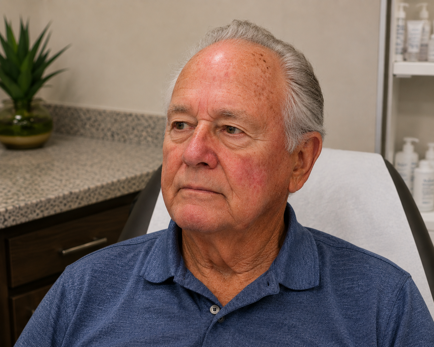
"My Forehead Has New Brown Spots, Red Dots, and Rough Bumps—Should I Be Worried About Skin Cancer?"
A 73-year-old man underwent a full-body skin examination that revealed benign moles, seborrheic keratoses, cherry angiomas, lentigines, and venous stasis. Learn why annual skin cancer screenings are essential for adults in Katy and Houston, Texas.
As we get older, it's common to notice new spots appearing on our skin. Some are rough and flaky, others are bright red, while some look like "stuck-on" brown growths. While many of these changes are completely harmless, distinguishing between benign age-related lesions and skin cancer isn't always easy.
Recently, a 73-year-old gentleman visited our dermatology clinic as a new patient for his annual full-body skin examination. He had noticed several skin lesions, including spots on his forehead, and wanted to be sure none of them were dangerous. After a comprehensive skin evaluation, our board-certified dermatologist identified several common age-related skin conditions—none of which represented skin cancer—but also reinforced why routine skin examinations remain one of the most important tools for early detection.
If you've ever wondered whether your new skin spots are normal aging or something more serious, this case highlights why regular skin checks are so valuable.
Why Annual Skin Exams Become More Important With Age
The risk of developing skin cancer increases steadily with age due to decades of cumulative ultraviolet (UV) exposure. Even people who rarely experienced severe sunburns often accumulate enough sun damage over time to develop new growths.
A full-body skin examination allows dermatologists to evaluate every area of the skin—including places patients cannot easily see themselves—to identify suspicious lesions before they become serious.
During this patient's comprehensive examination, every major skin surface was carefully evaluated using a dermatoscope, a specialized instrument that allows dermatologists to examine pigmented lesions with greater detail than the naked eye.
Benign Moles: Knowing What "Normal" Looks Like
Several of this patient's moles were identified as benign nevi.
Healthy moles generally have predictable characteristics:
Symmetrical shape
Smooth borders
Even color
Stable appearance over time
Although these moles required no treatment, the patient was encouraged to continue monthly self-skin examinations and monitor for changes using the familiar ABCDE warning signs of melanoma.
Sun Spots (Lentigines): Evidence of Years of Sun Exposure
The examination also revealed multiple solar lentigines, commonly called age spots or liver spots.
These flat tan or light brown patches develop after years of ultraviolet exposure and are especially common on the:
Forearms
Face
Shoulders
Hands
Unlike skin cancer, lentigines are completely benign.
While treatment is unnecessary, cosmetic improvement can often be achieved through:
Prescription retinoids
Chemical peels
Laser treatments
Medical-grade skincare
Daily sunscreen use
Cherry Angiomas: Those Bright Red "Blood Spots"
Like many adults over age 40, this patient also had several cherry angiomas on his back and abdomen.
Cherry angiomas are tiny collections of blood vessels that appear as bright red or cherry-colored bumps.
Although they may suddenly increase in number with age, they are completely benign and never become skin cancer.
Patients often choose removal only for cosmetic reasons using:
Laser therapy
Electrodessication
Otherwise, no treatment is necessary.
Seborrheic Keratoses: The Classic "Stuck-On" Growths
Several flat, waxy lesions were identified as seborrheic keratoses, one of the most common benign skin growths seen in older adults.
These growths frequently appear to be:
Brown
Gray
Tan
Slightly raised
Waxy or "stuck onto" the skin
Because one irritated lesion was located on the patient's forehead, a courtesy liquid nitrogen treatment (cryotherapy) was performed during the visit.
The patient was advised that if the lesion failed to resolve completely, additional evaluation could be performed during a follow-up appointment.
Venous Stasis: Skin Changes Caused by Poor Circulation
The examination also noted venous stasis changes affecting the lower legs.
Venous stasis occurs when weakened leg veins allow blood to pool in the lower extremities, leading to:
Swelling
Skin discoloration
Dryness
Itching
Increased risk of ulcers
The patient was already working with a vein specialist and was successfully losing weight, both of which can improve symptoms.
To further protect the skin, our dermatologist recommended:
Daily compression stockings
Regular moisturizers
Elevating the legs whenever possible
Patients should seek prompt medical attention if one leg suddenly becomes more swollen than the other, develops pain, ulceration, or signs of infection.
Why Sunscreen Is Still the Best Anti-Aging Treatment
Perhaps the most important recommendation during this visit involved daily sun protection.
Broad-spectrum sunscreen with SPF 30 or higher remains one of the simplest and most effective ways to reduce future sun damage.
Our dermatologists recommend:
Applying sunscreen 15 minutes before sun exposure
Reapplying every two hours while outdoors
Reapplying every 45–60 minutes after swimming or sweating
Using approximately one ounce (a shot-glass amount) to cover exposed skin
Wearing SPF lip balm
Using sun-protective clothing whenever practical
Daily sunscreen not only lowers the risk of skin cancer but also helps prevent additional sun spots, wrinkles, and premature aging.
The Bottom Line
Finding new skin spots as we age is incredibly common—but determining which ones are harmless and which deserve further evaluation requires expert examination.
In this patient's case, his annual skin examination identified multiple benign conditions, including moles, lentigines, cherry angiomas, seborrheic keratoses, and venous stasis changes. Although none required aggressive treatment, routine surveillance and ongoing sun protection remain essential to maintaining healthy skin.
Annual skin cancer screenings provide reassurance, establish a baseline for future comparison, and allow suspicious lesions to be detected long before they become dangerous.
Expert Skin Cancer Screenings in Katy & Houston, Texas
At Village Dermatology, our board-certified dermatologists perform comprehensive full-body skin examinations using advanced dermoscopy to detect skin cancer early while identifying and treating common benign skin conditions.
Whether you're concerned about a changing mole, rough growth, age spots, cherry angiomas, or simply want peace of mind through an annual skin cancer screening, our experienced team proudly serves patients throughout Katy, Houston, and surrounding Texas communities.
Protect your skin today by scheduling your annual full-body skin examination with Village Dermatology.
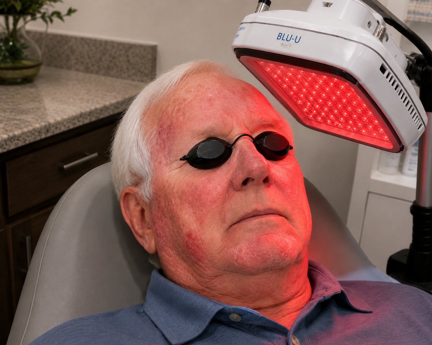
"I've Already Had My Pre-Cancerous Spots Frozen Off—Why Does My Dermatologist Recommend Blue Light Therapy Now?"
A 77-year-old patient learns why photodynamic therapy (PDT) with Ameluz® can be more effective than repeated cryotherapy for widespread actinic keratoses. Discover how Village Dermatology in Katy and Houston treats precancerous sun damage before it progresses to skin cancer.
Many patients are surprised when they return for a follow-up visit after having several rough, sun-damaged spots frozen with liquid nitrogen, only to hear their dermatologist recommend a completely different treatment.
A 77-year-old gentleman recently visited our dermatology clinic in Houston for follow-up of multiple actinic keratoses (AKs) affecting his face, nose, and lips. Although he had previously undergone cryotherapy, his board-certified dermatologist recommended photodynamic therapy (PDT) using Ameluz® and red light to better treat the remaining precancerous lesions.
If you've ever wondered why freezing isn't always enough—or why your dermatologist recommends blue or red light treatment for sun damage—this case explains the reasoning.
Understanding Actinic Keratoses
Actinic keratoses are rough, scaly patches that develop after years of cumulative ultraviolet (UV) exposure. They most commonly appear on areas that receive the greatest amount of sunlight, including:
Face
Nose
Lips
Ears
Bald scalp
Forearms
Hands
Although many AKs remain stable, they are considered precancerous lesions because a small percentage can progress into squamous cell carcinoma (SCC) if left untreated.
Because it is impossible to predict which lesion will become cancerous, dermatologists typically recommend treatment for all clinically apparent actinic keratoses.
Why Cryotherapy Isn't Always Enough
Cryotherapy (freezing individual spots with liquid nitrogen) remains one of the most effective treatments for isolated actinic keratoses.
However, many patients—especially those with years of sun exposure—develop field cancerization, meaning the surrounding skin contains numerous microscopic precancerous cells that are invisible to the naked eye.
In this patient's case, despite previous cryotherapy, there were still multiple erythematous, rough, hyperkeratotic lesions distributed across the:
Face
Nose
Lips
Rather than continuing to freeze each lesion individually, the dermatologist recommended treating the entire damaged field of skin.
What Is Photodynamic Therapy (PDT)?
Photodynamic therapy is an advanced, non-surgical treatment designed to eliminate both visible and early invisible actinic keratoses.
The treatment works in three steps:
Step 1: Preparing the Skin
Any thick, crusted lesions are gently curetted to allow better medication penetration.
Step 2: Applying Ameluz®
A photosensitizing medication called Ameluz® (aminolevulinic acid) is applied to the affected skin.
The medication is preferentially absorbed by abnormal sun-damaged cells while largely sparing healthy surrounding tissue.
Step 3: Red Light Activation
The treated skin is illuminated using a 635 nm red LED light for approximately 30 minutes.
When exposed to the specific wavelength of light, the medication creates oxygen radicals that selectively destroy damaged precancerous cells.
Why Treat the Entire Face?
One of PDT's biggest advantages is that it treats:
Visible actinic keratoses
Tiny developing lesions
Sun-damaged cells not yet visible
Instead of treating one lesion at a time, PDT addresses the entire field of damaged skin, lowering the burden of precancerous changes.
For many patients with widespread facial sun damage, this approach produces longer-lasting improvement than repeated spot treatments alone.
What Happens During Treatment?
Patients are often pleasantly surprised that the procedure is performed entirely in the dermatology office.
During this patient's visit:
Written informed consent was obtained.
Thick lesions were gently curetted.
The treatment areas were cleansed and prepared.
Protective eye shields were placed.
One tube of Ameluz® was applied to the face, nose, and lips.
The skin was illuminated with red light for 30 minutes.
The procedure required no incisions or stitches, allowing the patient to return home immediately afterward.
What Should Patients Expect After PDT?
Because PDT specifically targets damaged skin cells, it is normal to experience temporary reactions during healing.
Common side effects include:
Redness
Mild swelling
Burning sensation
Scabbing
Peeling
Temporary pigment changes
These reactions are expected and usually indicate that the medication is actively treating abnormal cells.
Why Avoid Sunlight After Treatment?
One of the most important instructions following PDT is strict avoidance of sunlight for at least 48 hours.
The photosensitizing medication remains temporarily active after treatment.
Even brief sun exposure can cause an exaggerated skin reaction.
Patients should:
Stay indoors whenever possible
Wear wide-brimmed hats
Use broad-spectrum sunscreen after the initial avoidance period
Avoid direct sunlight for the first two days
Following these instructions helps minimize discomfort and supports proper healing.
Can PDT Help Prevent Skin Cancer?
While no treatment completely eliminates future skin cancers, treating actinic keratoses significantly reduces the number of precancerous lesions that may eventually progress to squamous cell carcinoma.
Patients with:
Extensive sun damage
Multiple previous actinic keratoses
Prior skin cancers
Frequent cryotherapy treatments
often benefit from periodic field therapy like PDT as part of a long-term skin cancer prevention strategy.
Regular full-body skin examinations remain an essential part of ongoing care.
Expert Actinic Keratosis Treatment in Katy & Houston, Texas
If you have rough, scaly spots on your face, scalp, ears, or lips, don't assume they're simply part of aging.
At Village Dermatology, our board-certified dermatologists provide comprehensive evaluation and treatment for actinic keratoses, sun damage, and skin cancer prevention using advanced therapies including Ameluz® Photodynamic Therapy (PDT).
Whether you need cryotherapy, PDT, or routine skin cancer screenings, our team proudly serves patients throughout Houston, Katy, and surrounding Texas communities with personalized, evidence-based dermatologic care.
Schedule your skin evaluation today to protect your skin and catch precancerous changes before they become something more serious.
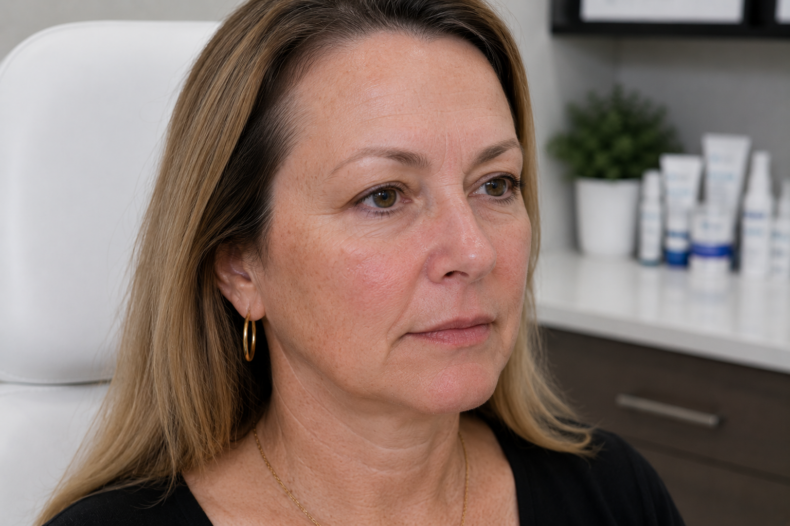
"I Have a History of Atypical Moles—Why Did My Dermatologist Just Biopsy a Bright Red Spot Instead?"
A 58-year-old patient with a history of atypical nevi visited Village Dermatology for a routine check, resulting in a shave biopsy of a suspicious red spot on her arm. Read our latest blog post to see why checking non-brown spots is critical to your health.
When you have a known history of atypical nevi (moles that look different than normal moles under a microscope), you tend to keep a very close eye on dark, asymmetrical, or changing brown spots. You schedule your routine skin checks expecting your doctor to focus entirely on those areas.
But during a recent visit to our dermatology clinic in Houston, a 58-year-old female patient experienced a common twist during her routine check. While she had a personal history of atypical moles, her exam revealed something she didn’t expect: a bright cherry-red bump on her upper arm that required an immediate medical biopsy.
Routine screenings are full of surprises because our skin changes as we age. Here is how our board-certified dermatologist in Houston evaluated her skin, why a bright red spot took priority, and what her visit teaches us about total skin health.
The Full-Body Exam: Tracking Atypical Moles and Beyond
Because this patient was proactive about her history of atypical nevi, she came in for a comprehensive, full-body skin examination. A thorough evaluation is critical for anyone with her skin profile, even though she had a reassuring list of "pertinent negatives"—meaning she had no personal or family history of melanoma or non-melanoma skin cancers.
During the head-to-toe exam, our clinical team noted several normal, benign signs of aging and sun exposure on her back and face, including:
Benign Nevi: Safe, symmetrical, common moles on her upper back.
Seborrheic Keratoses & Cherry Angiomas: Harmless, non-cancerous warty growths and standard red vascular spots on her spine and upper back.
Lentigines & Actinic Damage: Light tan sun spots and early signs of photoaging (textural thinning and sun damage) across her cheeks.
While these spots simply required standard sun-protection counseling, a completely different lesion on her left arm raised a clinical red flag.
Why a Bright Red Spot Required a Shave Biopsy
On the patient's left upper arm, our Houston dermatologist located a bright, cherry-red papule. While many small red spots are entirely harmless blood vessel growths (like the cherry angiomas on her back), this specific lesion had features that could mimic something more serious.
Our clinical differential diagnosis included a neoplasm of unspecified behavior versus squamous cell carcinoma (a common type of skin cancer that can sometimes present as an elevated, red, or crusting bump).
[Clinical Differential Diagnosis]
│
├─► Cherry Angioma (Benign vascular growth)
│
└─► Squamous Cell Carcinoma (Skin cancer mimicking a vascular spot)
│
└─► Action Required: Shave Biopsy for Lab Verification
To ensure her safety and obtain a definitive diagnosis, our skin care specialists in Katy Texas performed a quick, in-office biopsy by shave method:
Numbing: The area was prepped and numbed locally with a fast-acting anesthetic.
The Shave: Using a specialized, flexible blade (Dermablade), our provider gently shaved the lesion down to the upper layer of the dermis (the second layer of skin).
Healing: A specialized solution (Drysol) was applied to stop any minor bleeding, followed by protective petrolatum (Vaseline) and a sterile bandage.
The sample was safely sent to a dermatopathology lab for a formal microscopic evaluation to confirm exactly what the cells are.
The Ultimate Checklist for Sun Protection
Whether you are protecting atypical moles or trying to reverse photoaging and sun spots, your daily routine is your shield. Our Katy dermatology clinic provided the patient with the gold-standard protocol for preventing future skin damage:
The Shot Glass Rule: You need a full ounce of sunscreen—roughly the size of a shot glass—to adequately cover your body.
The 15-Minute Rule: Always apply your broad-spectrum mineral sunscreen (SPF 30 or higher) at least 15 minutes before stepping outside.
The Reapply Clock: Reapply every 2 hours normally, or every 45 to 60 minutes if you are sweating or swimming in the Texas heat.
Medical-Grade vs. OTC: Look for highly rated mineral formulas containing zinc oxide or titanium dioxide (such as EltaMD, ISDIN, or sensitive-skin over-the-counter options from CeraVe and La Roche-Posay).
Protect Your Skin: Schedule Your Annual Skin Check
This patient’s case is a prime example of why you cannot just monitor your own moles at home. It takes an expert eye and a clinical dermatoscope to spot the subtle differences between a harmless age spot and a lesion that requires a laboratory biopsy.
If you have a history of unique moles, or if you have spotted a new, changing, or bright red bump on your skin, do not wait. Schedule your comprehensive full-body examination with our dedicated team at Village Dermatology Houston or Katy today.
"Why Are My Lips Constantly Red, Peeling, and Burning No Matter How Much Lip Balm I Apply?"
A 51-year-old patient came to Village Dermatology with a severe, peeling lip rash that wouldn't heal. Discover how our Houston and Katy skin care specialists diagnosed allergic contact dermatitis, initiated a non-steroidal treatment plan, and used patch testing to find the root cause.
We have all dealt with chapped lips during a dry Texas breeze or a sudden weather shift. Usually, a quick swipe of lip balm does the trick. But what happens when your lips become severely red, dry, peeling, and painful, and nothing seems to soothe them?
A 51-year-old male patient recently visited our dermatology clinic in Houston experiencing this exact frustrating issue. He presented with a moderate to severe rash localized entirely on his lips. It wasn't just typical winter chapping; it was a persistent, uncomfortable inflammation that was actively disrupting his daily life.
When simple dryness turns into a severe, non-healing lip rash, it’s time to see a board-certified dermatologist in Houston or Katy to identify the underlying trigger. Here is what our clinical examination revealed and how we mapped out his path to relief.
Unmasking Lip Dermatitis: Is it an Allergy?
Upon evaluating the patient’s lips with a dermatoscope, our clinical team diagnosed him with allergic contact dermatitis of the lips (also known as lip dermatitis or cheilitis).
Allergic contact dermatitis is an inflammatory skin reaction that triggers a delayed hypersensitivity response. On the lips, this condition is incredibly tricky because it is frequently caused by the very products people use to treat dry skin. Common culprits include:
Fragrances, flavorings, or beeswax in popular lip balms.
Specific chemicals or foaming agents in toothpaste and mouthwashes.
Hidden allergens in shaving creams, foods, or skin care products.
To break the cycle of inflammation, our Houston dermatologist emphasized the absolute importance of stopping any chronic lip licking or skin picking, which strips the lips of their natural moisture barrier and perpetuates the painful rash.
The Investigation: Self-Elimination and Patch Testing
Finding the exact cause of allergic contact dermatitis requires a bit of detective work. Our skin care specialists in Katy Texas initiated a two-phase diagnostic strategy for the patient:
The Self-Elimination Trial: The patient was instructed to immediately strip his daily routine down to the basics. This meant eliminating all exposure to scented soaps, flavored lip products, essential oils, and complex cosmetics, switching instead to strictly hypoallergenic cleansers and moisturizers.
Comprehensive Patch Testing: Because the exact allergen was unknown, we scheduled the patient for patch testing at our Katy dermatology clinic. Patch testing is a specialized diagnostic tool where microscopic amounts of common allergens are safely applied to the skin (usually the back) and monitored over a few days to pinpoint exactly what is triggering the immune reaction.
The Non-Steroidal Treatment Plan for Sensitive Skin
Because the skin on the lips is thin and highly sensitive, utilizing long-term topical steroids can cause unwanted side effects like skin thinning. To safely calm his severe inflammation, our dermatologist in Katy TX prescribed tacrolimus 0.1% topical ointment to be applied twice daily.
Tacrolimus ointment is a highly effective, non-steroidal anti-inflammatory treatment. It targets the localized immune response to stop the redness, peeling, and burning without the risks associated with topical steroids.
How to Soothe Inflamed Lips at Home
If you are currently battling a mysterious, peeling lip rash, our medical team at Village Dermatology recommends taking these immediate steps before your appointment:
Switch to Hypoallergenic Cleansers: Avoid heavily fragranced facial washes. Opt for gentle, unscented options.
Simplify Your Lip Care: Stop using flavored, medicated, or cooling lip balms (especially those containing menthol, camphor, or fragrance). Stick to plain, pure white petrolatum.
Audit Your Toothpaste: Try switching to a basic, flavor-free toothpaste without heavy whitening agents or sodium lauryl sulfate (SLS), which are frequent irritants.
Get Relief for Your Skin Allergies at Village Dermatology
Allergic contact dermatitis can stubbornly persist for several weeks, and trying to guess the trigger on your own can lead to months of unnecessary discomfort.
If you are struggling with a persistent rash on your face, lips, or body, let our team provide the expert diagnostic care you deserve. Schedule an appointment at Village Dermatology Houston or Katy today to get a definitive answer and a personalized treatment plan.
"Could My Cancer Treatment Be Causing This Severe, Stubborn Adult Acne?"
A 67-year-old male patient visited Village Dermatology to treat severe facial acne while undergoing oncology care. Discover how our Houston and Katy skin care specialists adjusted his Isotretinoin treatment to achieve clear skin without interfering with his medical treatments.
When we think of acne, we usually picture teenagers navigating the hormonal shifts of puberty. But what happens when you are 67 years old and suddenly battling a severe breakout of inflammatory bumps and pustules on your face?
Recently, a 67-year-old male patient walked into our dermatology clinic in Houston for a follow-up appointment. He was dealing with a challenging case of severe adult acne that required aggressive intervention. What made his case unique—and highly complex—was that he was simultaneously undergoing oncology treatment.
Many patients do not realize that certain life-saving chemotherapy medications can heavily disrupt the skin, leading to severe, drug-induced breakouts or exacerbating underlying conditions like DSAP (Disseminated Superficial Actinic Porokeratosis, a rare type of sun-damage lesion).
Here is how our board-certified dermatologist in Houston navigated his care to safely clear his skin while coordinating with his oncology team.
The Challenge: Treating Acne During Chemotherapy
When this patient initially sought help from our skin care specialists in Katy Texas, his face showed deep inflammatory papules, pustules, and comedones (clogged pores). Because standard over-the-counter washes aren't enough for severe or medication-induced acne, he was started on standard protocol Isotretinoin (commonly known by its original brand name, Accutane).
At his 2-month follow-up, the patient reported great news: he was tolerating the medication incredibly well, with no major side effects other than manageable dry skin.
However, medical cases aren't always a straight line. The patient shared that his oncologist was switching his chemotherapy medication to a different drug. Our medical team took the time to explain that because these specific cancer therapies often fall into the same drug category, his facial breakouts might fluctuate. Managing skin health during cancer treatment requires a highly observant, expert dermatologist in Katy TX to balance powerful skin medications with oncological care.
Adjusting the Treatment Plan for Clear Skin
Because the patient’s face showed clear improvement but still had a few persistent, stubborn areas, our Houston dermatologist adjusted his treatment strategy to safely push past the plateau:
Increasing the Dosage: We increased his prescription to Isotretinoin 30 mg capsules (to be taken twice daily). Our target protocol is to reach a protective cumulative dose of 200–220 mg/kg to ensure long-term clearance.
Managing Body Spots: To address separate persistent areas on his body, we refilled a topical triamcinolone cream to calm localized inflammation daily.
Strict Safety Monitoring: Isotretinoin is an incredibly effective medication, but it requires strict medical supervision. We re-counseled the patient on vital safety measures, including mandatory monthly blood tests, avoiding night driving if vision changes occur, and postponing any elective surgeries for six months post-treatment.
The Daily Routine for Severe Adult Acne
Whether you are taking prescription medications or just trying to get a handle on late-stage breakouts, your daily habits matter. Our Katy dermatology clinic emphasizes a gentle, non-comedogenic (pore-friendly) foundation:
Non-Comedogenic Cleansers & Moisturizers: Powerful treatments like Isotretinoin cause significant dryness. You must use heavy, oil-free moisturizers and cleansers that repair the skin barrier without clogging your pores.
Broad-Spectrum Sunscreen SPF 30+: Acne medications make your skin highly sensitive to the intense Texas sun. Daily sun protection is non-negotiable to prevent permanent scarring and hyperpigmentation.
It can take up to 2 to 3 months to see a 60–80% improvement in severe acne. Patience, consistency, and expert medical guidance are the keys to success.
Get Expert Acne Care at Village Dermatology
Acne at age 67 is not just a cosmetic nuisance; it is a medical condition that deserves specialized, compassionate care—especially if you are navigating other health treatments like chemotherapy.
If you are searching for a dermatologist in Houston Texas who looks at your whole health history rather than just your skin surface, we are here to help. Schedule a comprehensive evaluation at Village Dermatology Houston or Katy today to start your journey toward comfortable, clear skin.
“Why Is My Ear Piercing Keloid Itching, and Can I Treat My Acne and Sweating While Pregnant?“
Wondering how to treat an itchy ear keloid or severe back acne while pregnant? Read a real patient case study from Village Dermatology Houston to learn about pregnancy-safe skin care, from topical clindamycin to postpartum keloid removal plans in Houston and Katy, Texas.
Pregnancy brings about incredible changes to your body, but it can also trigger unexpected skin changes. If you have noticed an old ear scar suddenly growing and itching, or if you are battling stubborn breakouts on your back while expecting, you are certainly not alone.
Many expecting mothers visit our clinics looking for safe, effective ways to manage complex skin conditions. In this case study, we look at a 26-year-old female patient who came to our office at eight months pregnant. She was dealing with an enlarging, itchy scar on her right ear, severe back breakouts, and a lifelong battle with excessive underarm sweating. Her visit highlights how we balance powerful dermatological care with the utmost safety for you and your baby.
Managing an Itchy, Enlarging Ear Keloid During Pregnancy
Our patient’s primary concern was a 1.2 cm firm, itchy growth on her right posterior earlobe that had been gradually enlarging over the past year. Using a specialized skin camera called a dermatoscope, our dermatologist in Houston diagnosed the growth as a keloid—an overgrowth of scar tissue that often forms after minor trauma, such as an ear piercing.
Keloids can become highly uncomfortable, causing persistent itching or pain. While there are several excellent options for keloid removal in Houston, pregnancy changes our timeline. The gold standard treatment for preventing a keloid from returning involves a surgical excision combined with a series of monthly corticosteroid injections for three consecutive months immediately following the procedure. Because the patient was heavily pregnant, our skin specialist in Katy and Houston recommended deferring the surgical removal and steroid injections until after she delivers her baby to ensure absolute safety for the infant.
Safe Pregnancy Solutions for Back Acne and Dark Spots
In addition to the ear keloid, the patient was dealing with a flare-up of acne across her upper back (the superior thoracic spine), which had also left behind dark spots, known medically as post-inflammatory hyperpigmentation (PIH).
When you are expecting, many standard acne medications (like high-strength retinoids) are completely off the table. However, you do not have to just suffer through the breakouts. Our Katy dermatology team designed a pregnancy-safe regimen to clear her skin:
Benzoyl Peroxide (BPO) Wash: A safe, over-the-counter antibacterial wash to clear out acne-causing bacteria from the pores on her back.
Topical Clindamycin 1% Gel: A prescription-strength, pregnancy-safe topical antibiotic applied daily to target deep inflammation without systemic risks.
Broad-Spectrum Sunscreen (SPF 30+): Hormonal fluctuations during pregnancy can cause dark spots and hyperpigmentation to worsen significantly with sun exposure. Applying a broad-spectrum sunscreen daily is essential to help those spots fade naturally over time.
We counseled the patient that while it typically takes two to three months to see a 60% to 80% improvement in acne, consistency now will protect her skin from developing permanent structural scars.
Dealing with Hyperhidrosis (Severe Sweating) While Expecting
The patient's final concern was a lifelong struggle with severe underarm sweating, an inherited genetic condition known as axillary hyperhidrosis. She was hoping for a clinical solution but found that common clinical options—including over-the-counter Certain Dri and prescription options like Qbrexza cloths or glycopyrrolate tablets—are not recommended for use during pregnancy.
Our medical team advised her to stick with standard, gentle antiperspirants for the remaining weeks of her pregnancy. Once she delivers and finishes breastfeeding, she can return to our clinic to explore highly effective hyperhidrosis treatment in Houston, which can include prescription topicals, oral medications, or even specialized Botox injections to temporarily block the sweat glands.
Are you pregnant and struggling with unexpected skin changes, stubborn acne, or an uncomfortable scar? You don’t have to guess which products are safe for your baby. Schedule a personalized consultation for safe, effective pregnancy dermatology solutions in Houston and Katy today.
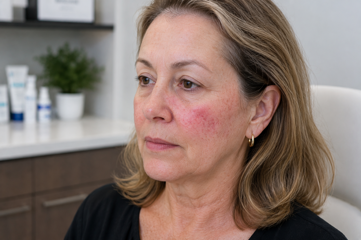
"Why Do I Have a Sudden Red Face Rash and Strange Skin Spots at 67?"
A 67-year-old patient came to Village Dermatology looking for answers to a stubborn red face rash and mysterious body lesions. Read our latest case study to find out how our Houston and Katy dermatologists diagnosed her rosacea, treated precancerous sun damage, and built the ultimate anti-aging skincare routine.
by: Dr. Ashley Baldree
As we gracefully age, our skin goes through a lot of transitions. It’s incredibly common to look in the mirror one day and notice a persistent flush on your cheeks, or feel a new rough patch on your skin that wasn't there before. If you are experiencing this, you are not alone.
Recently, a 67-year-old female patient visited our dermatology clinic in Houston seeking answers. She had two primary concerns: a red, moderate rash on her face that had been lingering for a month, and several asymptomatic skin lesions on her back and abdomen that she had carried for years.
Even though she had no personal or family history of skin cancer, she knew that getting a professional evaluation from a board-certified dermatologist in Houston was the safest choice for her peace of mind.
Here is what our expert team discovered during her comprehensive skin examination, and how we helped her restore her skin health.
Diagnosing the Red Face Rash: Managing Rosacea and Telangiectasia
Upon a detailed visual and dermatoscopic examination of her face, our Houston dermatologist identified the root cause of her red facial rash: rosacea accompanied by telangiectasia (scattered broken blood vessels) on her left cheek.
Rosacea is a chronic skin condition characterized by centrofacial redness, visible blood vessels, and sometimes small, pus-filled bumps. It can easily be mistaken for a standard adult acne breakout or a simple allergic reaction. Because rosacea is chronic, it requires specialized care from a skin care specialist in Katy Texas or Houston to keep flare-ups at bay.
The Customized Plan for Facial Redness:
Trigger Awareness: We counseled the patient on common lifestyle triggers that worsen flushing, including sun exposure, wind, hot temperatures, stress, alcohol, and spicy foods.
Cosmetic Solutions: To instantly minimize the appearance of redness, we recommended utilizing green-tinted moisturizers (such as Eucerin Redness Relief).
Advanced Laser Therapy: For a definitive solution to get rid of broken capillaries, we referred her to our cosmetic team to discuss VBEAM laser treatment, which target vascular redness at its source.
Evaluating the Body Lesions: Age Spots vs. Sun Damage
Next, our team evaluated the spots on her body. When you visit a trusted dermatologist in Katy TX, a full-body skin check is vital to differentiate between purely cosmetic growths and precancerous lesions.
Our exam revealed a mix of benign, age-related growths and sun-damaged areas:
Actinic Keratosis (AK): Our team identified rough, scaly patches on her left forehead, right hand, and lower leg. Unlike benign spots, actinic keratosis is a precancerous growth caused by cumulative sun damage. To protect her health, we treated these spots using liquid nitrogen (cryotherapy) to safely freeze away the damaged cells.
Seborrheic Keratoses & Cherry Angiomas: The patient also had several "stuck-on" waxy papules and small red vascular growths. These are completely benign, non-cancerous signs of aging skin. We performed a courtesy cosmetic liquid nitrogen treatment on an irritated seborrheic keratosis on her leg to alleviate discomfort.
Lentigines and Photoaging: Commonly known as age spots or sun spots, these flat, tan macules are the result of photoaging (sun damage).
The Gold Standard Prevention Regimen
The ultimate line of defense against both photoaging and worsening rosacea is strict sun protection. Our medical team prescribed a daily morning routine using a Vitamin C serum (like Vive-C) paired with a high-quality, broad-spectrum mineral sunscreen with an SPF of 30 or higher.
For patients shopping for the best medical-grade sun protection, our Katy dermatology clinic highly recommends brands like Elta MD or ISDIN. If you prefer over-the-counter options, ensure they are pure mineral formulas containing only zinc oxide or titanium dioxide, such as:
Supergoop Mineral Sheerscreen SPF 30
Eucerin Sensitive Mineral SPF 50
CeraVe Mineral Sunscreen SPF 50
Vanicream Sunscreen Broad Spectrum SPF 50
Neutrogena Sheer Zinc Face SPF 50
When Was Your Last Full-Body Skin Check?
This patient’s case highlights exactly why routine visits to a dermatologist in Houston Texas are so critical. While many of her spots were harmless signs of aging, she also required swift treatment for precancerous lesions she hadn't even realized were dangerous.
We scheduled her for a 1-year follow-up skin exam and encouraged her to perform monthly self-skin checks at home. If you notice a changing mole, a stubborn new red rash, or just want to reverse signs of sun damage, schedule an appointment with our dedicated team at Village Dermatology today!